PI 3-kinase p110α Polyclonal Antibody
- Catalog No.:YT3709
- Applications:IF;WB;IHC;ELISA
- Reactivity:Human;Mouse;Rat
- Target:
- PI 3-kinase p110α
- Fields:
- >>Inositol phosphate metabolism;>>Metabolic pathways;>>EGFR tyrosine kinase inhibitor resistance;>>Endocrine resistance;>>Platinum drug resistance;>>ErbB signaling pathway;>>Ras signaling pathway;>>Rap1 signaling pathway;>>cAMP signaling pathway;>>Chemokine signaling pathway;>>HIF-1 signaling pathway;>>FoxO signaling pathway;>>Phosphatidylinositol signaling system;>>Sphingolipid signaling pathway;>>Phospholipase D signaling pathway;>>Autophagy - animal;>>mTOR signaling pathway;>>PI3K-Akt signaling pathway;>>AMPK signaling pathway;>>Apoptosis;>>Longevity regulating pathway;>>Longevity regulating pathway - multiple species;>>Cellular senescence;>>Axon guidance;>>VEGF signaling pathway;>>Osteoclast differentiation;>>Focal adhesion;>>Signaling pathways regulating pluripotency of stem cells;>>Platelet activation;>>Neutrophil extracellular trap formation;>>Toll-like receptor signaling pathway;>>C-type lectin receptor signaling pathway;>>JAK-STAT signaling pathway;>>Natural killer cell mediat
- Gene Name:
- PIK3CA
- Protein Name:
- Phosphatidylinositol 4,5-bisphosphate 3-kinase catalytic subunit alpha isoform
- Human Gene Id:
- 5290
- Human Swiss Prot No:
- P42336
- Mouse Gene Id:
- 18706
- Mouse Swiss Prot No:
- P42337
- Immunogen:
- The antiserum was produced against synthesized peptide derived from human PI 3-kinase p110alpha. AA range:470-519
- Specificity:
- PI 3-kinase p110α Polyclonal Antibody detects endogenous levels of PI 3-kinase p110α protein.
- Formulation:
- Liquid in PBS containing 50% glycerol, 0.5% BSA and 0.02% sodium azide.
- Source:
- Polyclonal, Rabbit,IgG
- Dilution:
- IF 1:50-200 WB 1:500 - 1:2000. IHC 1:100 - 1:300. ELISA: 1:40000. Not yet tested in other applications.
- Purification:
- The antibody was affinity-purified from rabbit antiserum by affinity-chromatography using epitope-specific immunogen.
- Concentration:
- 1 mg/ml
- Storage Stability:
- -15°C to -25°C/1 year(Do not lower than -25°C)
- Other Name:
- PIK3CA;Phosphatidylinositol 4;5-bisphosphate 3-kinase catalytic subunit alpha isoform;PI3-kinase subunit alpha;PI3K-alpha;PI3Kalpha;PtdIns-3-kinase subunit alpha;Phosphatidylinositol 4,5-bisphosphate 3-kinase 110 kDa catalytic subunit
- Observed Band(KD):
- 110kD
- Background:
- Phosphatidylinositol 3-kinase is composed of an 85 kDa regulatory subunit and a 110 kDa catalytic subunit. The protein encoded by this gene represents the catalytic subunit, which uses ATP to phosphorylate PtdIns, PtdIns4P and PtdIns(4,5)P2. This gene has been found to be oncogenic and has been implicated in cervical cancers. A pseudogene of this gene has been defined on chromosome 22. [provided by RefSeq, Apr 2016],
- Function:
- catalytic activity:ATP + 1-phosphatidyl-1D-myo-inositol 4,5-bisphosphate = ADP + 1-phosphatidyl-1D-myo-inositol 3,4,5-trisphosphate.,disease:Defects in PIK3CA are associated with breast cancer [MIM:114480].,disease:Defects in PIK3CA are associated with colorectal cancer (CRC) [MIM:114500].,disease:Defects in PIK3CA are associated with ovarian cancer [MIM:167000]. Ovarian cancer is the leading cause of death from gynecologic malignancy. It is characterized by advanced presentation with loco-regional dissemination in the peritoneal cavity and the rare incidence of visceral metastases. These typical features relate to the biology of the disease, which is a principal determinant of outcome.,disease:Defects in PIK3CA may underlie hepatocellular carcinoma (HCC) [MIM:114550].,disease:PI3KCA mutations affecting exons 9 and 20 display gender-and tissue-specific patterns, thus suggesting that the
- Subcellular Location:
- intracellular,cytosol,plasma membrane,phosphatidylinositol 3-kinase complex,phosphatidylinositol 3-kinase complex, class IA,lamellipodium,
- Expression:
- Brain,Lung,
Over-Expression of Centromere Protein U Participates in the Malignant Neoplastic Progression of Breast Cancer. Frontiers in Oncology Front Oncol. 2021 Mar;0:350 WB Human 1 : 1000 Breast cancer tissues ,the paired adjacent histologically normal tissue T47D cell, MDA-MB-231 cell, SK-BR-3 cell, MCF-7 cell
HSP90 inhibitor AUY922 can reverse Fulvestrant induced feedback reaction in human breast cancer cells. CANCER SCIENCE 2017 May 19 WB Human MCF-7 cell,T47D cell
Deficiency in the short‐chain acyl‐CoA dehydrogenase protects mice against diet‐induced obesity and insulin resistance. FASEB JOURNAL Faseb J. 2019 Dec;33(12):13722-13733 WB Mouse liver hepatocytes
CD38 is involved in cell energy metabolism via activating the PI3K/AKT/mTOR signaling pathway in cervical cancer cells. INTERNATIONAL JOURNAL OF ONCOLOGY Int J Oncol. 2020 Jul;57(1):338-354 WB Human 1:1000 CaSki cell,Hela cell
Structural characterization of a pectic polysaccharide from Codonopsis pilosula and its immunomodulatory activities in vivo and in vitro. INTERNATIONAL JOURNAL OF BIOLOGICAL MACROMOLECULES Int J Biol Macromol. 2017 Nov;104:1359 WB Mouse Splenocytes
CD90 affects the biological behavior and energy metabolism level of gastric cancer cells by targeting the PI3K/AKT/HIF‑1α signaling pathway. Oncology Letters Oncol Lett. 2021 Mar;21(3):1-1 WB Human 1:1000 AGS cell
Buqi Huoxue Tongnao prescription protects against chronic cerebral hypoperfusion via regulating PI3K/AKT and LXRα/CYP7A1 signaling pathways PHYTOMEDICINE Yinhuang Gao WB Rat,Mouse 1:1000 hippocampus tissue BV2 cell
- June 19-2018
- WESTERN IMMUNOBLOTTING PROTOCOL
- June 19-2018
- IMMUNOHISTOCHEMISTRY-PARAFFIN PROTOCOL
- June 19-2018
- IMMUNOFLUORESCENCE PROTOCOL
- September 08-2020
- FLOW-CYTOMEYRT-PROTOCOL
- May 20-2022
- Cell-Based ELISA│解您多样本WB检测之困扰
- July 13-2018
- CELL-BASED-ELISA-PROTOCOL-FOR-ACETYL-PROTEIN
- July 13-2018
- CELL-BASED-ELISA-PROTOCOL-FOR-PHOSPHO-PROTEIN
- July 13-2018
- Antibody-FAQs
- Products Images
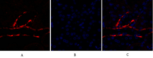
- Immunofluorescence analysis of rat-kidney tissue. 1,PI 3-kinase p110α Polyclonal Antibody(red) was diluted at 1:200(4°C,overnight). 2, Cy3 labled Secondary antibody was diluted at 1:300(room temperature, 50min).3, Picture B: DAPI(blue) 10min. Picture A:Target. Picture B: DAPI. Picture C: merge of A+B

- Immunofluorescence analysis of rat-kidney tissue. 1,PI 3-kinase p110α Polyclonal Antibody(red) was diluted at 1:200(4°C,overnight). 2, Cy3 labled Secondary antibody was diluted at 1:300(room temperature, 50min).3, Picture B: DAPI(blue) 10min. Picture A:Target. Picture B: DAPI. Picture C: merge of A+B
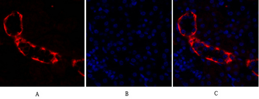
- Immunofluorescence analysis of mouse-kidney tissue. 1,PI 3-kinase p110α Polyclonal Antibody(red) was diluted at 1:200(4°C,overnight). 2, Cy3 labled Secondary antibody was diluted at 1:300(room temperature, 50min).3, Picture B: DAPI(blue) 10min. Picture A:Target. Picture B: DAPI. Picture C: merge of A+B
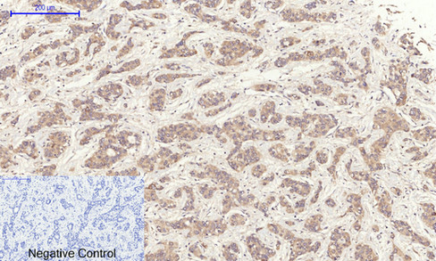
- Immunohistochemical analysis of paraffin-embedded Human-liver-cancer tissue. 1,PI 3-kinase p110α Polyclonal Antibody was diluted at 1:200(4°C,overnight). 2, Sodium citrate pH 6.0 was used for antibody retrieval(>98°C,20min). 3,Secondary antibody was diluted at 1:200(room tempeRature, 30min). Negative control was used by secondary antibody only.
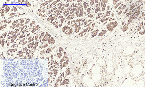
- Immunohistochemical analysis of paraffin-embedded Human-stomach-cancer tissue. 1,PI 3-kinase p110α Polyclonal Antibody was diluted at 1:200(4°C,overnight). 2, Sodium citrate pH 6.0 was used for antibody retrieval(>98°C,20min). 3,Secondary antibody was diluted at 1:200(room tempeRature, 30min). Negative control was used by secondary antibody only.
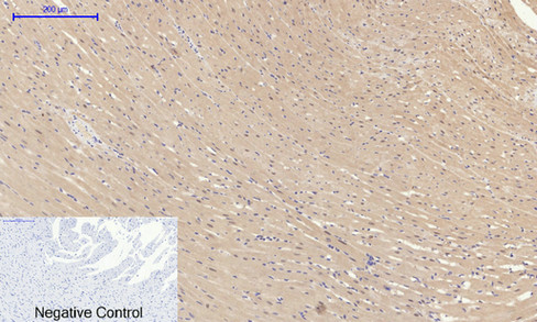
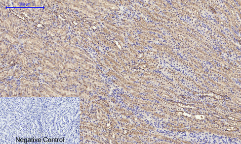
- Immunohistochemical analysis of paraffin-embedded Rat-heart tissue. 1,PI 3-kinase p110α Polyclonal Antibody was diluted at 1:200(4°C,overnight). 2, Sodium citrate pH 6.0 was used for antibody retrieval(>98°C,20min). 3,Secondary antibody was diluted at 1:200(room tempeRature, 30min). Negative control was used by secondary antibody only.
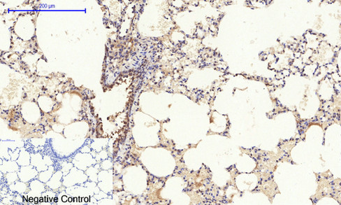
- Immunohistochemical analysis of paraffin-embedded Rat-lung tissue. 1,PI 3-kinase p110α Polyclonal Antibody was diluted at 1:200(4°C,overnight). 2, Sodium citrate pH 6.0 was used for antibody retrieval(>98°C,20min). 3,Secondary antibody was diluted at 1:200(room tempeRature, 30min). Negative control was used by secondary antibody only.
- Immunohistochemical analysis of paraffin-embedded Rat-kidney tissue. 1,PI 3-kinase p110α Polyclonal Antibody was diluted at 1:200(4°C,overnight). 2, Sodium citrate pH 6.0 was used for antibody retrieval(>98°C,20min). 3,Secondary antibody was diluted at 1:200(room tempeRature, 30min). Negative control was used by secondary antibody only.
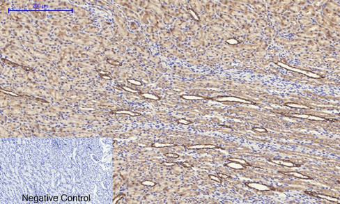
- Immunohistochemical analysis of paraffin-embedded Rat-spinal-cord tissue. 1,PI 3-kinase p110α Polyclonal Antibody was diluted at 1:200(4°C,overnight). 2, Sodium citrate pH 6.0 was used for antibody retrieval(>98°C,20min). 3,Secondary antibody was diluted at 1:200(room tempeRature, 30min). Negative control was used by secondary antibody only.
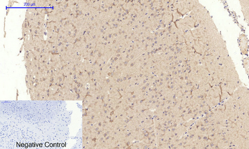
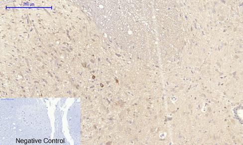
- Immunohistochemical analysis of paraffin-embedded Rat-brain tissue. 1,PI 3-kinase p110α Polyclonal Antibody was diluted at 1:200(4°C,overnight). 2, Sodium citrate pH 6.0 was used for antibody retrieval(>98°C,20min). 3,Secondary antibody was diluted at 1:200(room tempeRature, 30min). Negative control was used by secondary antibody only.
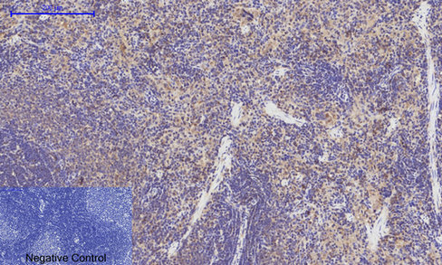
- Immunohistochemical analysis of paraffin-embedded Rat-spleen tissue. 1,PI 3-kinase p110α Polyclonal Antibody was diluted at 1:200(4°C,overnight). 2, Sodium citrate pH 6.0 was used for antibody retrieval(>98°C,20min). 3,Secondary antibody was diluted at 1:200(room tempeRature, 30min). Negative control was used by secondary antibody only.

- Immunohistochemical analysis of paraffin-embedded Mouse-heart tissue. 1,PI 3-kinase p110α Polyclonal Antibody was diluted at 1:200(4°C,overnight). 2, Sodium citrate pH 6.0 was used for antibody retrieval(>98°C,20min). 3,Secondary antibody was diluted at 1:200(room tempeRature, 30min). Negative control was used by secondary antibody only.

- Immunohistochemical analysis of paraffin-embedded Mouse-liver tissue. 1,PI 3-kinase p110α Polyclonal Antibody was diluted at 1:200(4°C,overnight). 2, Sodium citrate pH 6.0 was used for antibody retrieval(>98°C,20min). 3,Secondary antibody was diluted at 1:200(room tempeRature, 30min). Negative control was used by secondary antibody only.
- Immunohistochemical analysis of paraffin-embedded Mouse-kidney tissue. 1,PI 3-kinase p110α Polyclonal Antibody was diluted at 1:200(4°C,overnight). 2, Sodium citrate pH 6.0 was used for antibody retrieval(>98°C,20min). 3,Secondary antibody was diluted at 1:200(room tempeRature, 30min). Negative control was used by secondary antibody only.
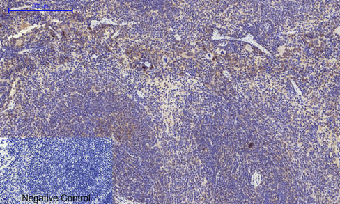
- Immunohistochemical analysis of paraffin-embedded Mouse-spleen tissue. 1,PI 3-kinase p110α Polyclonal Antibody was diluted at 1:200(4°C,overnight). 2, Sodium citrate pH 6.0 was used for antibody retrieval(>98°C,20min). 3,Secondary antibody was diluted at 1:200(room tempeRature, 30min). Negative control was used by secondary antibody only.
.jpg)
- Western Blot analysis of HELA cells using PI 3-kinase p110α Polyclonal Antibody diluted at 1:2000

- Western blot analysis of lysate from mouse liver, using PI 3-kinase p110α antibody.


