PEDF Polyclonal Antibody
- Catalog No.:YT3658
- Applications:IHC;IF;WB;ELISA
- Reactivity:Human;Mouse;Rat
- Target:
- PEDF
- Fields:
- >>Wnt signaling pathway
- Gene Name:
- SERPINF1
- Protein Name:
- Pigment epithelium-derived factor
- Human Gene Id:
- 5176
- Human Swiss Prot No:
- P36955
- Mouse Gene Id:
- 20317
- Mouse Swiss Prot No:
- P97298
- Immunogen:
- The antiserum was produced against synthesized peptide derived from human PEDF. AA range:258-307
- Specificity:
- PEDF Polyclonal Antibody detects endogenous levels of PEDF protein.
- Formulation:
- Liquid in PBS containing 50% glycerol, 0.5% BSA and 0.02% sodium azide.
- Source:
- Polyclonal, Rabbit,IgG
- Dilution:
- WB 1:500 - 1:2000. IHC: 1:100-300 ELISA: 1:20000. IF 1:100-300 Not yet tested in other applications.
- Purification:
- The antibody was affinity-purified from rabbit antiserum by affinity-chromatography using epitope-specific immunogen.
- Concentration:
- 1 mg/ml
- Storage Stability:
- -15°C to -25°C/1 year(Do not lower than -25°C)
- Other Name:
- SERPINF1;PEDF;PIG35;Pigment epithelium-derived factor;PEDF;Cell proliferation-inducing gene 35 protein;EPC-1;Serpin F1
- Observed Band(KD):
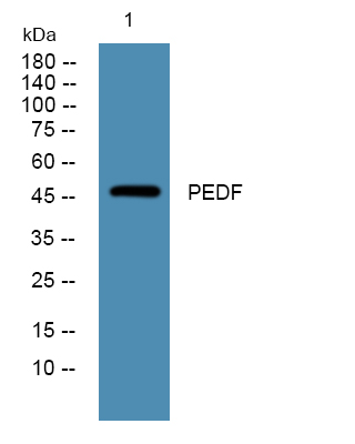
- 46kD
- Background:
- This gene encodes a member of the serpin family that does not display the serine protease inhibitory activity shown by many of the other serpin proteins. The encoded protein is secreted and strongly inhibits angiogenesis. In addition, this protein is a neurotrophic factor involved in neuronal differentiation in retinoblastoma cells. Mutations in this gene were found in individuals with osteogenesis imperfecta, type VI. [provided by RefSeq, Aug 2016],
- Function:
- developmental stage:Expressed in quiescent cells.,domain:The N-terminal (AA 44-121) exhibits neurite outgrowth-inducing activity. The C-terminal exposed loop (AA 382-418) is essential for serpin activity.,function:Neurotrophic protein; induces extensive neuronal differentiation in retinoblastoma cells. Potent inhibitor of angiogenesis. As it does not undergo the S (stressed) to R (relaxed) conformational transition characteristic of active serpins, it exhibits no serine protease inhibitory activity.,PTM:The N-terminus is blocked. Extracellular phosphorylation enhances antiangiogenic activity.,similarity:Belongs to the serpin family.,subcellular location:Enriched in stage I melanosomes.,tissue specificity:Retinal pigment epithelial cells and blood plasma.,
- Subcellular Location:
- Secreted . Melanosome . Enriched in stage I melanosomes.
- Expression:
- Retinal pigment epithelial cells and blood plasma.
The Role of RAD6B and PEDF in Retinal Degeneration. NEUROSCIENCE Neuroscience. 2022 Jan;480:19 WB,IHC Mouse 1:10000,1:200 Eyeball,Retina
- June 19-2018
- WESTERN IMMUNOBLOTTING PROTOCOL
- June 19-2018
- IMMUNOHISTOCHEMISTRY-PARAFFIN PROTOCOL
- June 19-2018
- IMMUNOFLUORESCENCE PROTOCOL
- September 08-2020
- FLOW-CYTOMEYRT-PROTOCOL
- May 20-2022
- Cell-Based ELISA│解您多样本WB检测之困扰
- July 13-2018
- CELL-BASED-ELISA-PROTOCOL-FOR-ACETYL-PROTEIN
- July 13-2018
- CELL-BASED-ELISA-PROTOCOL-FOR-PHOSPHO-PROTEIN
- July 13-2018
- Antibody-FAQs
- Products Images

- Immunofluorescence analysis of A549. 1,primary Antibody was diluted at 1:200(4°C overnight). 2, Goat Anti Rabbit IgG (H&L) - Alexa Fluor 488 Secondary antibody was diluted at 1:1000(room temperature, 50min).3, Picture B: DAPI(blue) 10min.

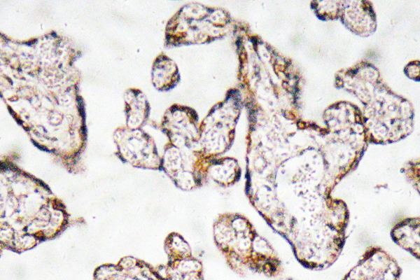
- Immunohistochemistry analysis of PEDF antibody in paraffin-embedded human prostate carcinoma tissue.

- Western blot analysis of lysates from A431 cells, primary antibody was diluted at 1:1000, 4°over night


