p53AIP1 Polyclonal Antibody
- Catalog No.:YT3542
- Applications:IHC;IF;ELISA
- Reactivity:Human;Rat;Mouse;
- Target:
- p53AIP1
- Fields:
- >>p53 signaling pathway;>>Apoptosis
- Gene Name:
- TP53AIP1
- Protein Name:
- p53-regulated apoptosis-inducing protein 1
- Human Gene Id:
- 63970
- Human Swiss Prot No:
- Q9HCN2
- Immunogen:
- The antiserum was produced against synthesized peptide derived from human TPIP1. AA range:75-124
- Specificity:
- p53AIP1 Polyclonal Antibody detects endogenous levels of p53AIP1 protein.
- Formulation:
- Liquid in PBS containing 50% glycerol, 0.5% BSA and 0.02% sodium azide.
- Source:
- Polyclonal, Rabbit,IgG
- Dilution:
- IHC 1:100 - 1:300. IF 1:200 - 1:1000. ELISA: 1:5000. Not yet tested in other applications.
- Purification:
- The antibody was affinity-purified from rabbit antiserum by affinity-chromatography using epitope-specific immunogen.
- Concentration:
- 1 mg/ml
- Storage Stability:
- -15°C to -25°C/1 year(Do not lower than -25°C)
- Other Name:
- TP53AIP1;p53-regulated apoptosis-inducing protein 1;p53AIP1
- Molecular Weight(Da):
- 13kD
- Background:
- This gene is specifically expressed in the thymus, and encodes a protein that is localized to the mitochondrion. The expression of this gene is inducible by p53, and it is thought to play an important role in mediating p53-dependent apoptosis. Alternatively spliced transcript variants encoding different isoforms have been described for this gene. [provided by RefSeq, Oct 2011],
- Function:
- function:May play an important role in mediating p53/TP53-dependent apoptosis.,induction:By p53/TP53.,tissue specificity:Only found to be expressed in thymus.,
- Subcellular Location:
- Mitochondrion .
- Expression:
- Only found to be expressed in thymus.
- June 19-2018
- WESTERN IMMUNOBLOTTING PROTOCOL
- June 19-2018
- IMMUNOHISTOCHEMISTRY-PARAFFIN PROTOCOL
- June 19-2018
- IMMUNOFLUORESCENCE PROTOCOL
- September 08-2020
- FLOW-CYTOMEYRT-PROTOCOL
- May 20-2022
- Cell-Based ELISA│解您多样本WB检测之困扰
- July 13-2018
- CELL-BASED-ELISA-PROTOCOL-FOR-ACETYL-PROTEIN
- July 13-2018
- CELL-BASED-ELISA-PROTOCOL-FOR-PHOSPHO-PROTEIN
- July 13-2018
- Antibody-FAQs
- Products Images
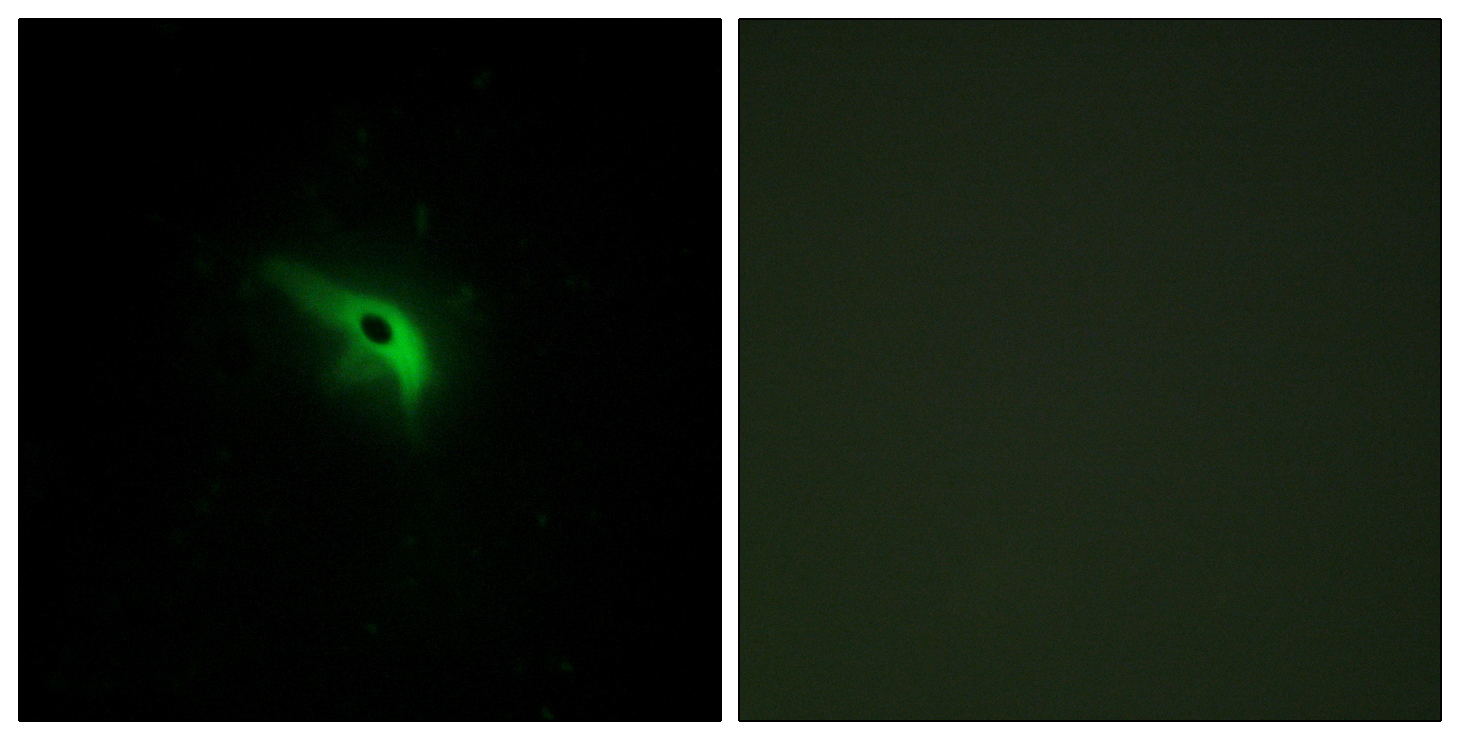
- Immunofluorescence analysis of A549 cells, using TPIP1 Antibody. The picture on the right is blocked with the synthesized peptide.
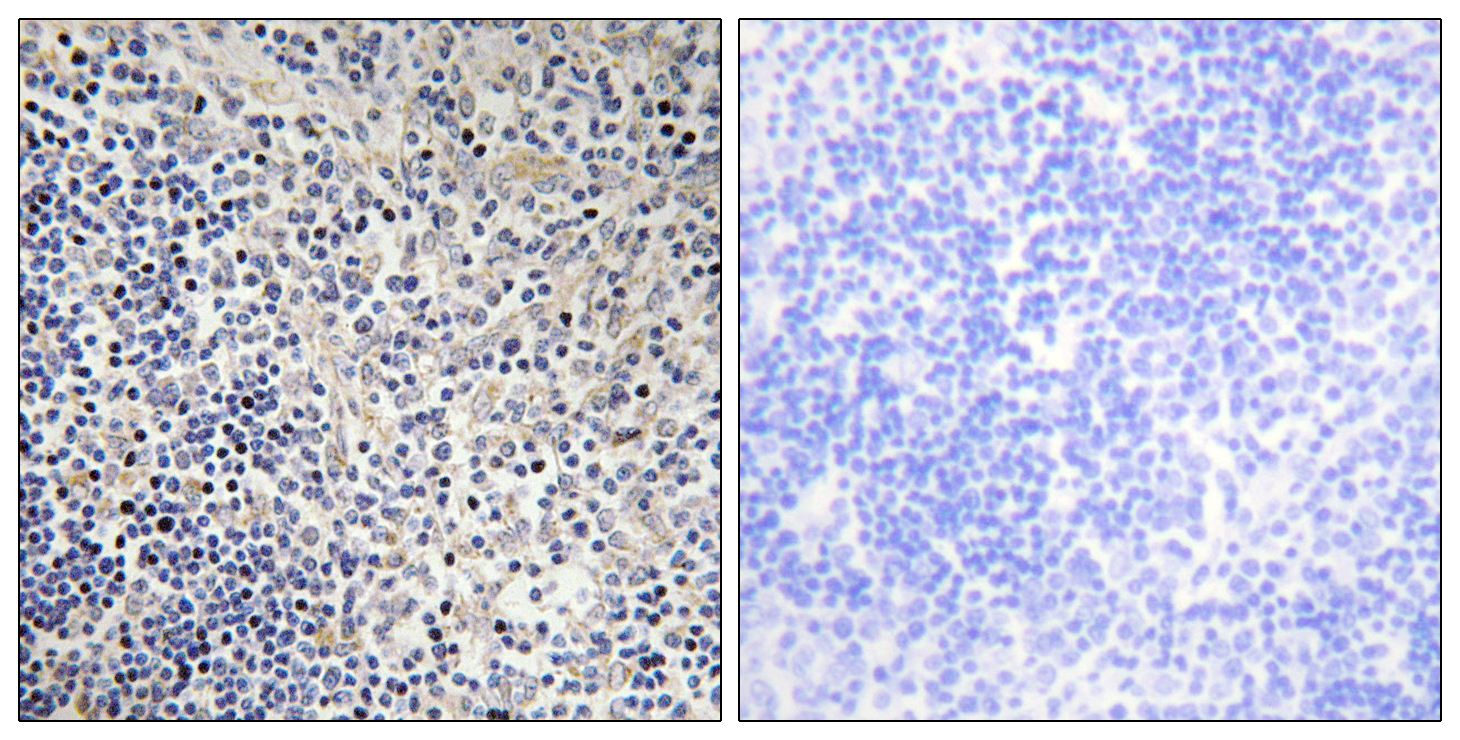
- Immunohistochemistry analysis of paraffin-embedded human thymus gland tissue, using TPIP1 Antibody. The picture on the right is blocked with the synthesized peptide.


