p35 Polyclonal Antibody
- Catalog No.:YT3510
- Applications:WB;IHC;IF;ELISA
- Reactivity:Human;Mouse;Rat
- Target:
- p35
- Fields:
- >>Alzheimer disease;>>Pathways of neurodegeneration - multiple diseases;>>Cocaine addiction
- Gene Name:
- CDK5R1
- Protein Name:
- Cyclin-dependent kinase 5 activator 1
- Human Gene Id:
- 8851
- Human Swiss Prot No:
- Q15078
- Mouse Gene Id:
- 12569
- Mouse Swiss Prot No:
- P61809
- Rat Gene Id:
- 116671
- Rat Swiss Prot No:
- P61810
- Immunogen:
- The antiserum was produced against synthesized peptide derived from human CDK5R1. AA range:11-60
- Specificity:
- p35 Polyclonal Antibody detects endogenous levels of p35 protein.
- Formulation:
- Liquid in PBS containing 50% glycerol, 0.5% BSA and 0.02% sodium azide.
- Source:
- Polyclonal, Rabbit,IgG
- Dilution:
- WB 1:500 - 1:2000. IHC 1:100 - 1:300. IF 1:200 - 1:1000. ELISA: 1:20000. Not yet tested in other applications.
- Purification:
- The antibody was affinity-purified from rabbit antiserum by affinity-chromatography using epitope-specific immunogen.
- Concentration:
- 1 mg/ml
- Storage Stability:
- -15°C to -25°C/1 year(Do not lower than -25°C)
- Other Name:
- CDK5R1;CDK5R;NCK5A;Cyclin-dependent kinase 5 activator 1;CDK5 activator 1;Cyclin-dependent kinase 5 regulatory subunit 1;TPKII regulatory subunit
- Observed Band(KD):
- 38kD
- Background:
- The protein encoded by this gene (p35) is a neuron-specific activator of cyclin-dependent kinase 5 (CDK5); the activation of CDK5 is required for proper development of the central nervous system. The p35 form of this protein is proteolytically cleaved by calpain, generating a p25 form. The cleavage of p35 into p25 results in relocalization of the protein from the cell periphery to nuclear and perinuclear regions. P25 deregulates CDK5 activity by prolonging its activation and changing its cellular location. The p25 form accumulates in the brain neurons of patients with Alzheimer's disease. This accumulation correlates with an increase in CDK5 kinase activity, and may lead to aberrantly phosphorylated forms of the microtubule-associated protein tau, which contributes to Alzheimer's disease. [provided by RefSeq, Jul 2008],
- Function:
- disease:Cleavage of p35 to p25 may be involved in the pathogenesis of Alzheimer disease. The p25 form accumulates in neurons in the brain of patients with Alzheimer disease, but not in normal brain. This accumulation correlates with an increase in CDK5 kinase activity. Application of amyloid beta peptide A-beta(1-42) induced the conversion of p35 to p25 in primary cortical neurons. Expression of the p25/Cdk5 complex in cultured primary neurons induces cytoskeletal disruption, morphological degeneration and apoptosis.,function:p35 is a neuron specific activator of CDK5. The complex p35/CDK5 is required for neurite outgrowth and cortical lamination. Activator of TPKII.,PTM:Probably myristoylated. The Gly-2-Ala mutant is absent of the cell periphery, suggesting that a proper myristoylation signal is essential for the proper distribution of p35.,PTM:The p35 form is proteolytically cleaved by
- Subcellular Location:
- [Cyclin-dependent kinase 5 activator 1, p35]: Cell membrane ; Lipid-anchor ; Cytoplasmic side . Cell projection, neuron projection . In the primary cortical neurons, p35 is present in the peripheries and nerve terminals. .; [Cyclin-dependent kinase 5 activator 1, p25]: Nucleus . Cytoplasm, perinuclear region . Perikaryon . The conversion of p35 to p25 relocalizes the protein from the cell periphery to the cytoplasm, in nuclear and perinuclear regions (PubMed:18507738). In the primary cortical neurons, p25 is primarily concentrated in the cell soma and is largely absent from neurites (PubMed:18507738). .
- Expression:
- Brain and neuron specific.
- June 19-2018
- WESTERN IMMUNOBLOTTING PROTOCOL
- June 19-2018
- IMMUNOHISTOCHEMISTRY-PARAFFIN PROTOCOL
- June 19-2018
- IMMUNOFLUORESCENCE PROTOCOL
- September 08-2020
- FLOW-CYTOMEYRT-PROTOCOL
- May 20-2022
- Cell-Based ELISA│解您多样本WB检测之困扰
- July 13-2018
- CELL-BASED-ELISA-PROTOCOL-FOR-ACETYL-PROTEIN
- July 13-2018
- CELL-BASED-ELISA-PROTOCOL-FOR-PHOSPHO-PROTEIN
- July 13-2018
- Antibody-FAQs
- Products Images
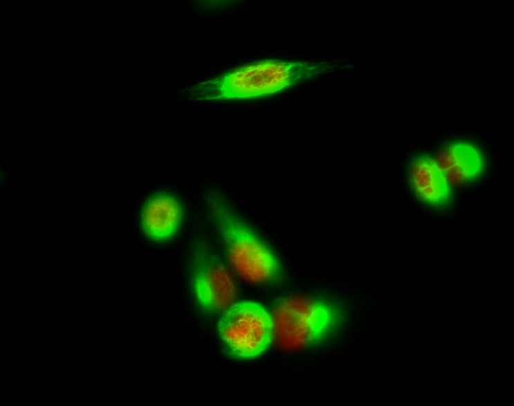
- Immunofluorescence analysis of Hela cell. 1,p35 Polyclonal Antibody(red) was diluted at 1:200(4° overnight). Caspase 9 Monoclonal Antibody(3-20)(green) was diluted at 1:200(4° overnight). 2, Goat Anti Rabbit Alexa Fluor 594 Catalog:RS3611 was diluted at 1:1000(room temperature, 50min). Goat Anti Mouse Alexa Fluor 488 Catalog:RS3208 was diluted at 1:1000(room temperature, 50min).
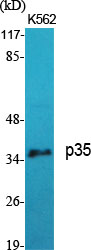
- Western Blot analysis of various cells using p35 Polyclonal Antibody diluted at 1:1000
.jpg)
- Western Blot analysis of HELA cells using p35 Polyclonal Antibody diluted at 1:1000
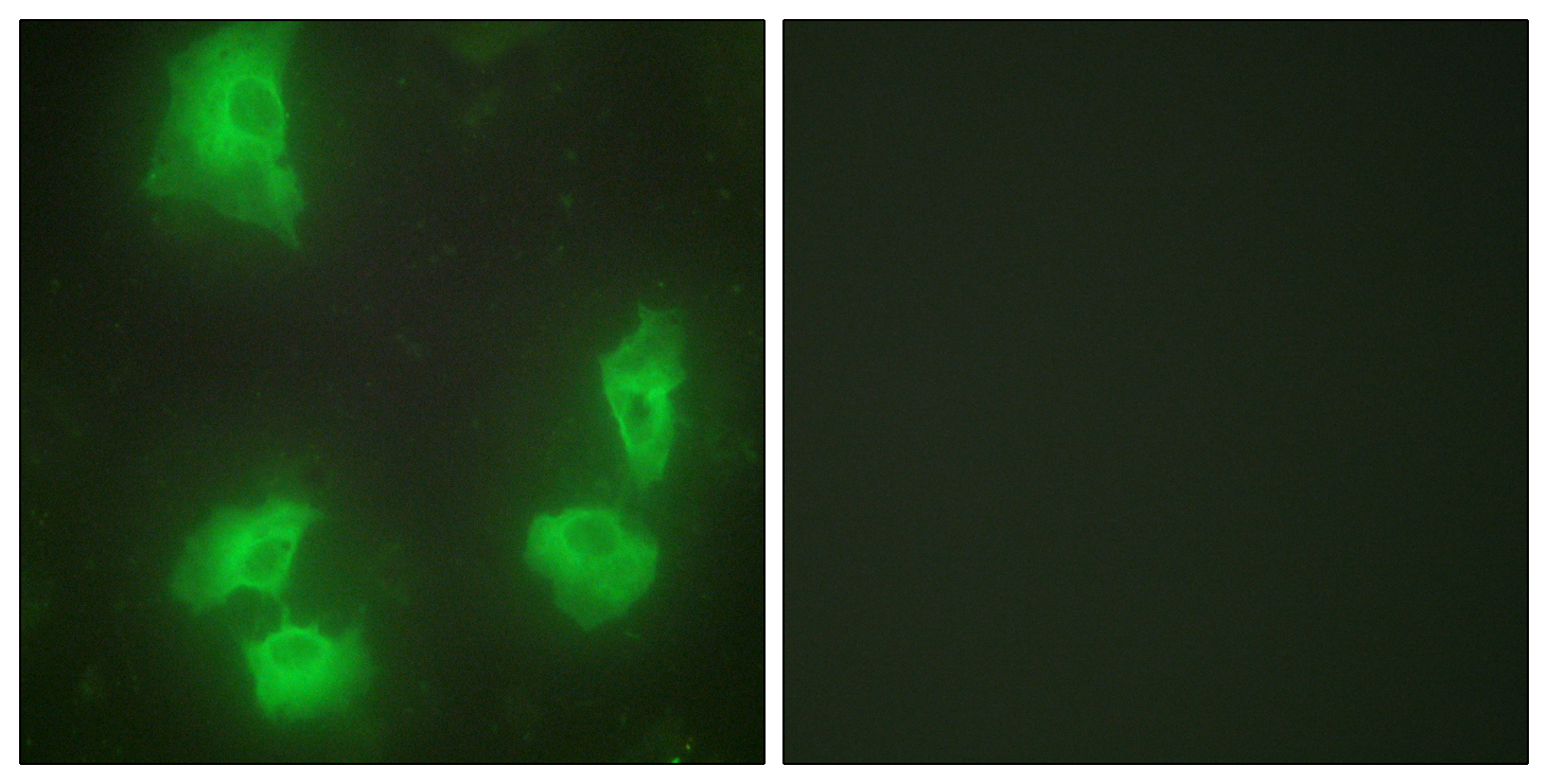
- Immunofluorescence analysis of HeLa cells, using CDK5R1 Antibody. The picture on the right is blocked with the synthesized peptide.
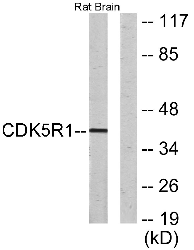
- Western blot analysis of lysates from rat brain cells, using CDK5R1 Antibody. The lane on the right is blocked with the synthesized peptide.

- Immunohistochemical analysis of paraffin-embedded human small intestinal carcinoma tissue. 1,primary Antibody was diluted at 1:200(4° overnight). 2, Sodium citrate pH 6.0 was used for antigen retrieval(>98°C,20min). 3,Secondary antibody was diluted at 1:200


