Nephrocystin-5 Polyclonal Antibody
- Catalog No.:YT3038
- Applications:WB;IHC;IF;ELISA
- Reactivity:Human;Mouse
- Target:
- Nephrocystin-5
- Gene Name:
- IQCB1
- Protein Name:
- IQ calmodulin-binding motif-containing protein 1
- Human Gene Id:
- 9657
- Human Swiss Prot No:
- Q15051
- Mouse Swiss Prot No:
- Q8BP00
- Immunogen:
- The antiserum was produced against synthesized peptide derived from human IQCB1. AA range:431-480
- Specificity:
- Nephrocystin-5 Polyclonal Antibody detects endogenous levels of Nephrocystin-5 protein.
- Formulation:
- Liquid in PBS containing 50% glycerol, 0.5% BSA and 0.02% sodium azide.
- Source:
- Polyclonal, Rabbit,IgG
- Dilution:
- WB 1:500 - 1:2000. IHC 1:100 - 1:300. ELISA: 1:40000.. IF 1:50-200
- Purification:
- The antibody was affinity-purified from rabbit antiserum by affinity-chromatography using epitope-specific immunogen.
- Concentration:
- 1 mg/ml
- Storage Stability:
- -15°C to -25°C/1 year(Do not lower than -25°C)
- Other Name:
- IQCB1;KIAA0036;NPHP5;OK/SW-cl.85;IQ calmodulin-binding motif-containing protein 1;Nephrocystin-5;p53 and DNA damage-regulated IQ motif protein;PIQ
- Observed Band(KD):
- 69kD
- Background:
- This gene encodes a nephrocystin protein that interacts with calmodulin and the retinitis pigmentosa GTPase regulator protein. The encoded protein has a central coiled-coil region and two calmodulin-binding IQ domains. It is localized to the primary cilia of renal epithelial cells and connecting cilia of photoreceptor cells. The protein is thought to play a role in ciliary function. Defects in this gene result in Senior-Loken syndrome type 5. Alternative splicing results in multiple transcript variants. A pseudogene of this gene is found on chromosome 6. [provided by RefSeq, Jan 2016],
- Function:
- disease:Defects in IQCB1 are the cause of Senior-Loken syndrome type 5 (SLSN5) [MIM:609254]. SLSN is a renal-retinal disorder, characterized by progressive wasting of the filtering unit of the kidney (nephronophthisis), with or without medullary cystic renal disease, and progressive eye disease. Typically this disorder becomes apparent during the first year of life.,similarity:Contains 4 IQ domains.,subunit:Interacts with calmodulin.,tissue specificity:Ubiquitously expressed in fetal and adult tissues. Localized to the outer segments and connecting cilia of photoreceptor cells.,
- Subcellular Location:
- Cytoplasm, cytoskeleton, microtubule organizing center, centrosome . Cytoplasm, cytoskeleton, microtubule organizing center, centrosome, centriole . Localization to the centrosome depends on the interaction with CEP290/NPHP6.
- Expression:
- Ubiquitously expressed in fetal and adult tissues. Localized to the outer segments and connecting cilia of photoreceptor cells. Up-regulated in a number of primary colorectal and gastric tumors.
- June 19-2018
- WESTERN IMMUNOBLOTTING PROTOCOL
- June 19-2018
- IMMUNOHISTOCHEMISTRY-PARAFFIN PROTOCOL
- June 19-2018
- IMMUNOFLUORESCENCE PROTOCOL
- September 08-2020
- FLOW-CYTOMEYRT-PROTOCOL
- May 20-2022
- Cell-Based ELISA│解您多样本WB检测之困扰
- July 13-2018
- CELL-BASED-ELISA-PROTOCOL-FOR-ACETYL-PROTEIN
- July 13-2018
- CELL-BASED-ELISA-PROTOCOL-FOR-PHOSPHO-PROTEIN
- July 13-2018
- Antibody-FAQs
- Products Images

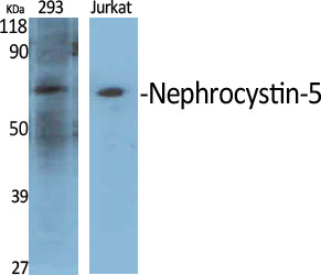
- Western Blot analysis of various cells using Nephrocystin-5 Polyclonal Antibody diluted at 1:500
.jpg)
- Western Blot analysis of SH-SY5Y cells using Nephrocystin-5 Polyclonal Antibody diluted at 1:500

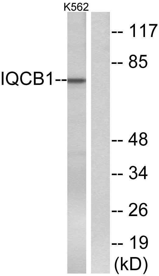
- Western blot analysis of lysates from K562 cells, using IQCB1 Antibody. The lane on the right is blocked with the synthesized peptide.

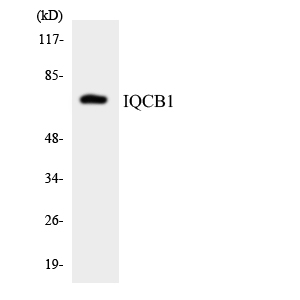
- Western blot analysis of the lysates from HepG2 cells using IQCB1 antibody.

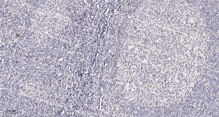
- Immunohistochemical analysis of paraffin-embedded human tonsil. 1, Antibody was diluted at 1:200(4° overnight). 2, Tris-EDTA,pH9.0 was used for antigen retrieval. 3,Secondary antibody was diluted at 1:200(room temperature, 30min).


