N33 Polyclonal Antibody
- Catalog No.:YT2961
- Applications:WB;IHC;IF;ELISA
- Reactivity:Human;Mouse;Rat
- Target:
- N33
- Fields:
- >>N-Glycan biosynthesis;>>Various types of N-glycan biosynthesis;>>Metabolic pathways;>>Protein processing in endoplasmic reticulum
- Gene Name:
- TUSC3
- Protein Name:
- Tumor suppressor candidate 3
- Human Gene Id:
- 7991
- Human Swiss Prot No:
- Q13454
- Mouse Gene Id:
- 80286
- Mouse Swiss Prot No:
- Q8BTV1
- Immunogen:
- The antiserum was produced against synthesized peptide derived from human TUSC3. AA range:131-180
- Specificity:
- N33 Polyclonal Antibody detects endogenous levels of N33 protein.
- Formulation:
- Liquid in PBS containing 50% glycerol, 0.5% BSA and 0.02% sodium azide.
- Source:
- Polyclonal, Rabbit,IgG
- Dilution:
- WB 1:500 - 1:2000. IHC 1:100 - 1:300. IF 1:200 - 1:1000. ELISA: 1:10000. Not yet tested in other applications.
- Purification:
- The antibody was affinity-purified from rabbit antiserum by affinity-chromatography using epitope-specific immunogen.
- Concentration:
- 1 mg/ml
- Storage Stability:
- -15°C to -25°C/1 year(Do not lower than -25°C)
- Other Name:
- TUSC3;N33;Tumor suppressor candidate 3;Magnesium uptake/transporter TUSC3;Protein N33
- Observed Band(KD):
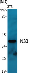
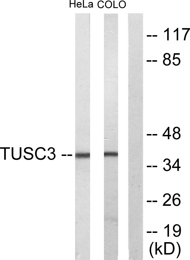
- 39kD
- Background:
- This gene is a candidate tumor suppressor gene. It is located within a homozygously deleted region of a metastatic prostate cancer. The gene is expressed in most nonlymphoid human tissues including prostate, lung, liver, and colon. Expression was also detected in many epithelial tumor cell lines. Two transcript variants encoding distinct isoforms have been identified for this gene. [provided by RefSeq, Jul 2008],
- Function:
- disease:Defects in TUSC3 are the cause of mental retardation non-syndromic autosomal recessive type 7 (MRT7) [MIM:611093]. Mental retardation is characterized by significantly sub-average general intellectual functioning associated with impairments in adaptative behavior and manifested during the developmental period. Non-syndromic mental retardation patients do not manifest other clinical signs.,function:May be involved in N-glycosylation through its association with N-oligosaccharyl transferase.,similarity:Belongs to the OST3/OST6 family.,subunit:Weakly associates with the oligosaccharyl transferase (OST) complex which contains at least RPN1/ribophorin I, RPN2/ribophorin II, OST48, DAD1, and either STT3A or STT3B.,tissue specificity:Expressed in most non-lymphoid cells and tissues examined, including prostate, lung, liver, colon, heart, kidney and pancreas.,
- Subcellular Location:
- Endoplasmic reticulum membrane ; Multi-pass membrane protein .
- Expression:
- Expressed in most non-lymphoid cells and tissues examined, including prostate, lung, liver, colon, heart, kidney and pancreas.
- June 19-2018
- WESTERN IMMUNOBLOTTING PROTOCOL
- June 19-2018
- IMMUNOHISTOCHEMISTRY-PARAFFIN PROTOCOL
- June 19-2018
- IMMUNOFLUORESCENCE PROTOCOL
- September 08-2020
- FLOW-CYTOMEYRT-PROTOCOL
- May 20-2022
- Cell-Based ELISA│解您多样本WB检测之困扰
- July 13-2018
- CELL-BASED-ELISA-PROTOCOL-FOR-ACETYL-PROTEIN
- July 13-2018
- CELL-BASED-ELISA-PROTOCOL-FOR-PHOSPHO-PROTEIN
- July 13-2018
- Antibody-FAQs
- Products Images

- Western Blot analysis of various cells using N33 Polyclonal Antibody
.jpg)
- Western Blot analysis of HeLa cells using N33 Polyclonal Antibody

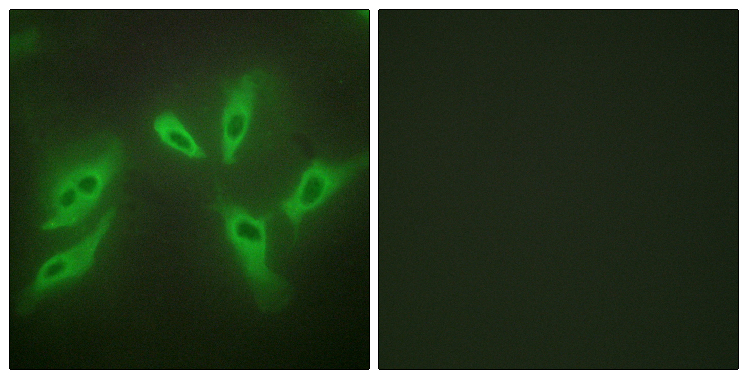
- Immunofluorescence analysis of HeLa cells using TUSC3 Antibody. The picture on the right is blocked with the synthesized peptide.

- Western blot analysis of lysates from COLO205 and HeLa cells, using TUSC3 Antibody. The lane on the right is blocked with the synthesized peptide.

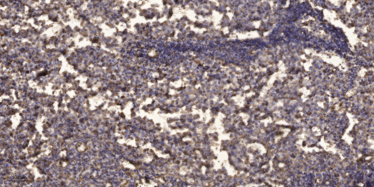
- Immunohistochemical analysis of paraffin-embedded human Squamous cell carcinoma of lung. 1, Antibody was diluted at 1:200(4° overnight). 2, Tris-EDTA,pH9.0 was used for antigen retrieval. 3,Secondary antibody was diluted at 1:200(room temperature, 45min).


