Myp Polyclonal Antibody
- Catalog No.:YT2953
- Applications:WB;IHC;IF;ELISA
- Reactivity:Human;Rat;Mouse;
- Target:
- Myp
- Gene Name:
- NOL3
- Protein Name:
- Nucleolar protein 3
- Human Gene Id:
- 8996
- Human Swiss Prot No:
- O60936-2
- Immunogen:
- The antiserum was produced against synthesized peptide derived from human ARC. AA range:159-208
- Specificity:
- Myp Polyclonal Antibody detects endogenous levels of Myp protein.
- Formulation:
- Liquid in PBS containing 50% glycerol, 0.5% BSA and 0.02% sodium azide.
- Source:
- Polyclonal, Rabbit,IgG
- Dilution:
- WB 1:500 - 1:2000. IHC 1:100 - 1:300. IF 1:200 - 1:1000. ELISA: 1:20000. Not yet tested in other applications.
- Purification:
- The antibody was affinity-purified from rabbit antiserum by affinity-chromatography using epitope-specific immunogen.
- Concentration:
- 1 mg/ml
- Storage Stability:
- -15°C to -25°C/1 year(Do not lower than -25°C)
- Other Name:
- Myp;NOL3;Nop30;Nucleolar protein 3;apoptosis repressor ARC;apoptosis repressor with CARD;apoptosis repressor with caspase recruitment domain (CARD);muscle-enriched cytoplasmic protein;nucleolar protein of 30 kDa
- Observed Band(KD):
- 28kD
- Background:
- NOL3 encodes an anti-apoptotic protein nucleolar protein 3 that has been shown to down-regulate the enzyme activities of caspase 2, caspase 8 and tumor protein p53. Multiple transcript variants encoding different isoforms have been found for NOL3.
- Subcellular Location:
- [Isoform 1]: Nucleus, nucleolus . The SR-rich C-terminus mediates nuclear localization. .; [Isoform 3]: Cytoplasm .; [Isoform 2]: Cytoplasm . Mitochondrion . Sarcoplasmic reticulum . Membrane ; Lipid-anchor . Phosphorylation at Thr-149 results in translocation to mitochondria. Colocalized with mitochondria in response to oxidative stress. .
- Expression:
- Highly expressed in heart and skeletal muscle. Detected at low levels in placenta, liver, kidney and pancreas.
- June 19-2018
- WESTERN IMMUNOBLOTTING PROTOCOL
- June 19-2018
- IMMUNOHISTOCHEMISTRY-PARAFFIN PROTOCOL
- June 19-2018
- IMMUNOFLUORESCENCE PROTOCOL
- September 08-2020
- FLOW-CYTOMEYRT-PROTOCOL
- May 20-2022
- Cell-Based ELISA│解您多样本WB检测之困扰
- July 13-2018
- CELL-BASED-ELISA-PROTOCOL-FOR-ACETYL-PROTEIN
- July 13-2018
- CELL-BASED-ELISA-PROTOCOL-FOR-PHOSPHO-PROTEIN
- July 13-2018
- Antibody-FAQs
- Products Images

- Western Blot analysis of various cells using Myp Polyclonal Antibody
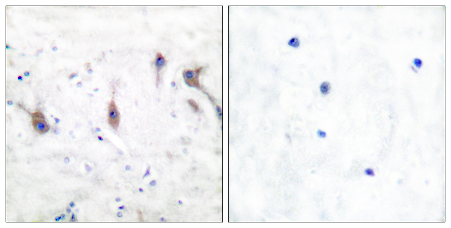
- Immunohistochemistry analysis of paraffin-embedded human brain tissue, using ARC Antibody. The picture on the right is blocked with the synthesized peptide.
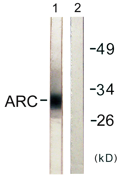
- Western blot analysis of lysates from HeLa cells, using ARC Antibody. The lane on the right is blocked with the synthesized peptide.

- Western Blot analysis of lane1 mouse-brain, lane2 mouse-kidney, lane3 Hela. land4 MCF7, lane5 293T, lane6 mouse-muscle using primary antibody at 1:1000 dilution 4°C, overnight. Secondary antibody(catalog#:RS23920) was diluted at 1:10000 25°C,1.5hours


