MMP-13 Polyclonal Antibody
- Catalog No.:YT2796
- Applications:WB;IHC;IF;ELISA
- Reactivity:Human;Rat;Mouse;
- Target:
- MMP-13
- Fields:
- >>IL-17 signaling pathway;>>Relaxin signaling pathway;>>Parathyroid hormone synthesis, secretion and action
- Gene Name:
- MMP13
- Protein Name:
- Collagenase 3
- Human Gene Id:
- 4322
- Human Swiss Prot No:
- P45452
- Mouse Swiss Prot No:
- P33435
- Immunogen:
- The antiserum was produced against synthesized peptide derived from human MMP-13. AA range:10-59
- Specificity:
- MMP-13 Polyclonal Antibody detects endogenous levels of MMP-13 protein.
- Formulation:
- Liquid in PBS containing 50% glycerol, 0.5% BSA and 0.02% sodium azide.
- Source:
- Polyclonal, Rabbit,IgG
- Dilution:
- WB 1:500 - 1:2000. IHC: 1:100-300 ELISA: 1:20000. IF 1:100-300 Not yet tested in other applications.
- Purification:
- The antibody was affinity-purified from rabbit antiserum by affinity-chromatography using epitope-specific immunogen.
- Concentration:
- 1 mg/ml
- Storage Stability:
- -15°C to -25°C/1 year(Do not lower than -25°C)
- Other Name:
- MMP13;Collagenase 3;Matrix metalloproteinase-13;MMP-13
- Observed Band(KD):
- 60kD
- Background:
- This gene encodes a member of the peptidase M10 family of matrix metalloproteinases (MMPs). Proteins in this family are involved in the breakdown of extracellular matrix in normal physiological processes, such as embryonic development, reproduction, and tissue remodeling, as well as in disease processes, such as arthritis and metastasis. The encoded preproprotein is proteolytically processed to generate the mature protease. This protease cleaves type II collagen more efficiently than types I and III. It may be involved in articular cartilage turnover and cartilage pathophysiology associated with osteoarthritis. Mutations in this gene are associated with metaphyseal anadysplasia. This gene is part of a cluster of MMP genes on chromosome 11. [provided by RefSeq, Jan 2016],
- Function:
- cofactor:Binds 2 zinc ions per subunit.,cofactor:Binds 4 calcium ions per subunit.,disease:Defects in MMP13 are the cause of spondyloepimetaphyseal dysplasia type 2 (SEMD2) [MIM:602111]; also known as spondyloepimetaphyseal dysplasia type Missouri. SEMDs are a heterogeneous group of skeletal disorders characterized by defective growth and modeling of the spine and long bones. The SEMDs are distinguished from the spondylometaphyseal dysplasias and the spondyloepiphyseal dysplasias by the combined involvement of the epiphyses and metaphyses. The 3 disorders have malformations of the vertebrae in common.,domain:The conserved cysteine present in the cysteine-switch motif binds the catalytic zinc ion, thus inhibiting the enzyme. The dissociation of the cysteine from the zinc ion upon the activation-peptide release activates the enzyme.,function:Degrades collagen type I. Does not act on gelati
- Subcellular Location:
- Secreted, extracellular space, extracellular matrix . Secreted .
- Expression:
- Detected in fetal cartilage and calvaria, in chondrocytes of hypertrophic cartilage in vertebrae and in the dorsal end of ribs undergoing ossification, as well as in osteoblasts and periosteal cells below the inner periosteal region of ossified ribs. Detected in chondrocytes from in joint cartilage that have been treated with TNF and IL1B, but not in untreated chondrocytes. Detected in T lymphocytes. Detected in breast carcinoma tissue.
17β-Estradiol alleviates intervertebral disc degeneration by inhibiting NF-κB signal pathway. LIFE SCIENCES Life Sci. 2021 Nov;284:119874 IHC Rat 1:200 Interverbal disc (IVD)
The regulatory role and mechanism of lncTUG1 on cartilage apoptosis and inflammation in osteoarthritis. Xiao-Yu Chen WB Human 1:1000 C28/I2 cell
Early Continuous Passive Motion and Needle-Knife Therapy Alleviate Knee Motor Dysfunction Effects After Internal Fixation of TPFs. Li Jia IHC Rabbit 1:100 ligamentous tissue
Cucurbitacin E reduces IL-1β-induced inflammation and cartilage degeneration by inhibiting the PI3K/Akt pathway in osteoarthritic chondrocytes Journal of Translational Medicine Wang Lin WB,IF,IHC Mouse,Human 1:1000,1:200,1:200 articular cartilage Chondrocytes
Koumine inhibits IL-1β-induced chondrocyte inflammation and ameliorates extracellular matrix degradation in osteoarthritic cartilage through activation of PINK1/Parkin-mediated mitochondrial autophagy BIOMEDICINE & PHARMACOTHERAPY Xiangyi Kong IHC,WB Rat 1:100 articular cartilage tissue RCCS-1 cell
Phillyrin reduces ROS production to alleviate the progression of intervertebral disc degeneration by inhibiting NF-κB pathway Journal of Orthopaedic Surgery and Research Chen Enming WB Rat 1:1000 nucleus pulposus cell (NPC)
- June 19-2018
- WESTERN IMMUNOBLOTTING PROTOCOL
- June 19-2018
- IMMUNOHISTOCHEMISTRY-PARAFFIN PROTOCOL
- June 19-2018
- IMMUNOFLUORESCENCE PROTOCOL
- September 08-2020
- FLOW-CYTOMEYRT-PROTOCOL
- May 20-2022
- Cell-Based ELISA│解您多样本WB检测之困扰
- July 13-2018
- CELL-BASED-ELISA-PROTOCOL-FOR-ACETYL-PROTEIN
- July 13-2018
- CELL-BASED-ELISA-PROTOCOL-FOR-PHOSPHO-PROTEIN
- July 13-2018
- Antibody-FAQs
- Products Images
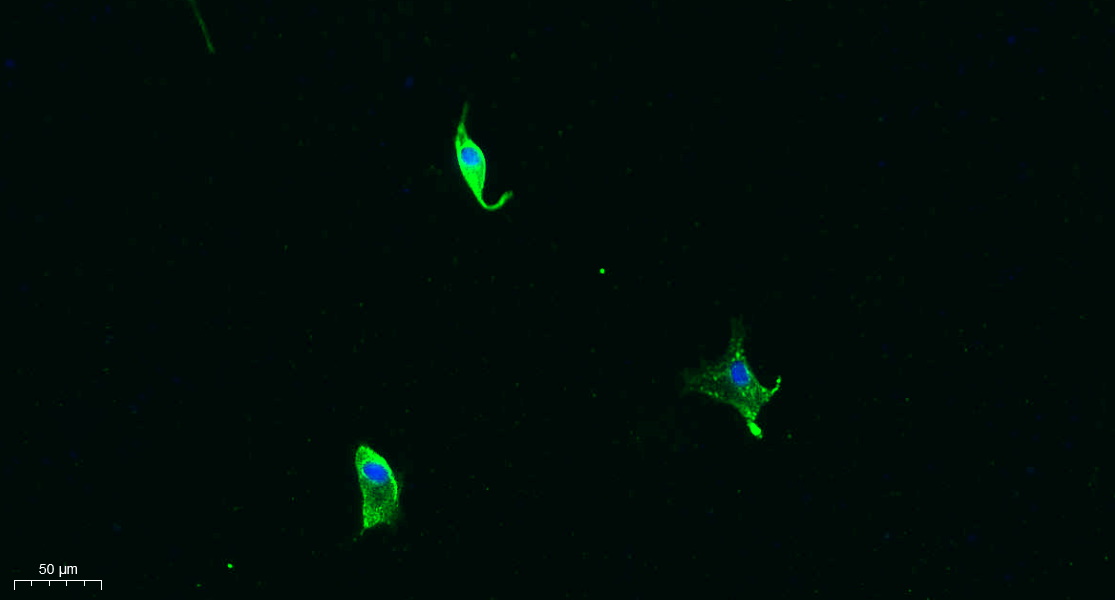
- Immunofluorescence analysis of A549. 1,primary Antibody was diluted at 1:200(4°C overnight). 2, Goat Anti Rabbit IgG (H&L) - Alexa Fluor 488 Secondary antibody was diluted at 1:1000(room temperature, 50min).3, Picture B: DAPI(blue) 10min.

- Western Blot analysis of various cells using MMP-13 Polyclonal Antibody diluted at 1:500
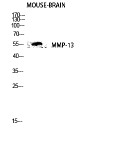
- Western blot analysis of mouse-brain lysis using MMP-13 antibody. Antibody was diluted at 1:500
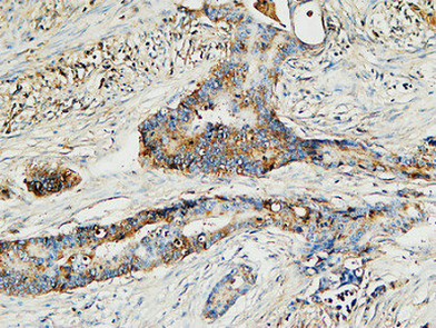
- Immunohistochemical analysis of paraffin-embedded Human Colon cancer. 1, Antibody was diluted at 1:100(4° overnight). 2, High-pressure and temperature EDTA, pH8.0 was used for antigen retrieval. 3,Secondary antibody was diluted at 1:200(room temperature, 30min).
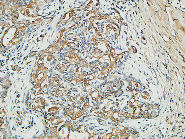
- Immunohistochemical analysis of paraffin-embedded Human Mammary cancer. 1, Antibody was diluted at 1:200(4° overnight). 2, High-pressure and temperature EDTA, pH8.0 was used for antigen retrieval. 3,Secondary antibody was diluted at 1:200(room temperature, 30min).

- Immunofluorescence analysis of HepG2 cells, using MMP-13 Antibody. The picture on the right is blocked with the synthesized peptide.
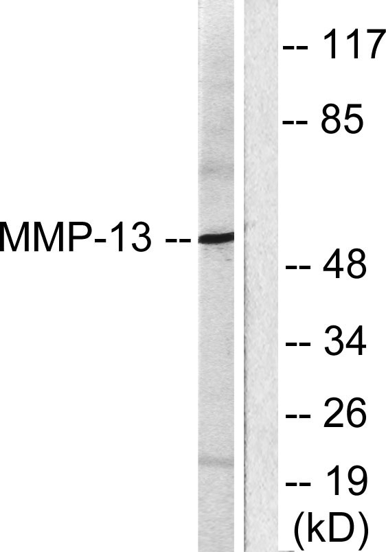
- Western blot analysis of lysates from LOVO cells, using MMP-13 Antibody. The lane on the right is blocked with the synthesized peptide.


