Mena Polyclonal Antibody
- Catalog No.:YT2731
- Applications:WB;IHC;IF;ELISA
- Reactivity:Human;Mouse;Rat
- Target:
- Mena
- Fields:
- >>Rap1 signaling pathway;>>Axon guidance;>>Regulation of actin cytoskeleton
- Gene Name:
- ENAH
- Protein Name:
- Protein enabled homolog
- Human Gene Id:
- 55740
- Human Swiss Prot No:
- Q8N8S7
- Mouse Gene Id:
- 13800
- Mouse Swiss Prot No:
- Q03173
- Immunogen:
- The antiserum was produced against synthesized peptide derived from human ENAH. AA range:472-521
- Specificity:
- Mena Polyclonal Antibody detects endogenous levels of Mena protein.
- Formulation:
- Liquid in PBS containing 50% glycerol, 0.5% BSA and 0.02% sodium azide.
- Source:
- Polyclonal, Rabbit,IgG
- Dilution:
- WB 1:500 - 1:2000. IHC 1:100 - 1:300. ELISA: 1:10000.. IF 1:50-200
- Purification:
- The antibody was affinity-purified from rabbit antiserum by affinity-chromatography using epitope-specific immunogen.
- Concentration:
- 1 mg/ml
- Storage Stability:
- -15°C to -25°C/1 year(Do not lower than -25°C)
- Other Name:
- ENAH;MENA;Protein enabled homolog
- Observed Band(KD):
- 67kD
- Background:
- This gene encodes a member of the enabled/ vasodilator-stimulated phosphoprotein. Members of this gene family are involved in actin-based motility. This protein is involved in regulating the assembly of actin filaments and modulates cell adhesion and motility. Alternate splice variants of this gene have been correlated with tumor invasiveness in certain tissues and these variants may serve as prognostic markers. A pseudogene of this gene is found on chromosome 3. [provided by RefSeq, Sep 2016],
- Function:
- domain:The EVH2 domain is comprised of 3 regions. Block A is a thymosin-like domain required for G-actin binding. The KLKR motif within this block is essential for the G-actin binding and for actin polymerization. Block B is required for F-actin binding and subcellular location, and Block C for tetramerization.,function:Ena/VASP proteins are actin-associated proteins involved in a range of processes dependent on cytoskeleton remodeling and cell polarity such as axon guidance and lamellipodial and filopodial dynamics in migrating cells. ENAH induces the formation of F-actin rich outgrowths in fibroblasts. Acts syngeristically with BAIAP2-alpha and downstream of NTN1 to promote filipodia formation. Required for the actin-based mobility of Listeria monocytogenes.,PTM:NTN1-induced PKA phosphorylation on Ser-265 directly parallels the formation of filopodial protrusions.,PTM:Phosphorylated up
- Subcellular Location:
- Cytoplasm. Cytoplasm, cytoskeleton . Cell projection, lamellipodium . Cell projection, filopodium . Cell junction, synapse . Cell junction, focal adhesion. Targeted to the leading edge of lamellipodia and filopodia by MRL family members. Colocalizes at filopodial tips with a number of other proteins including vinculin and zyxlin. Colocalizes with N-WASP at the leading edge. Colocalizes with GPHN and PFN at synapses (By similarity). .
- Expression:
- Expressed in myoepithelia of parotid, breast, bronchial glands and sweat glands. Expressed in colon-rectum muscolaris mucosae epithelium, pancreas acinar ductal epithelium, endometrium epithelium, prostate fibromuscolar stroma and placenta vascular media. Overexpressed in a majority of breast cancer cell lines and primary breast tumor lesions.
- June 19-2018
- WESTERN IMMUNOBLOTTING PROTOCOL
- June 19-2018
- IMMUNOHISTOCHEMISTRY-PARAFFIN PROTOCOL
- June 19-2018
- IMMUNOFLUORESCENCE PROTOCOL
- September 08-2020
- FLOW-CYTOMEYRT-PROTOCOL
- May 20-2022
- Cell-Based ELISA│解您多样本WB检测之困扰
- July 13-2018
- CELL-BASED-ELISA-PROTOCOL-FOR-ACETYL-PROTEIN
- July 13-2018
- CELL-BASED-ELISA-PROTOCOL-FOR-PHOSPHO-PROTEIN
- July 13-2018
- Antibody-FAQs
- Products Images

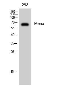
- Western Blot analysis of 293 cells using Mena Polyclonal Antibody diluted at 1:2000

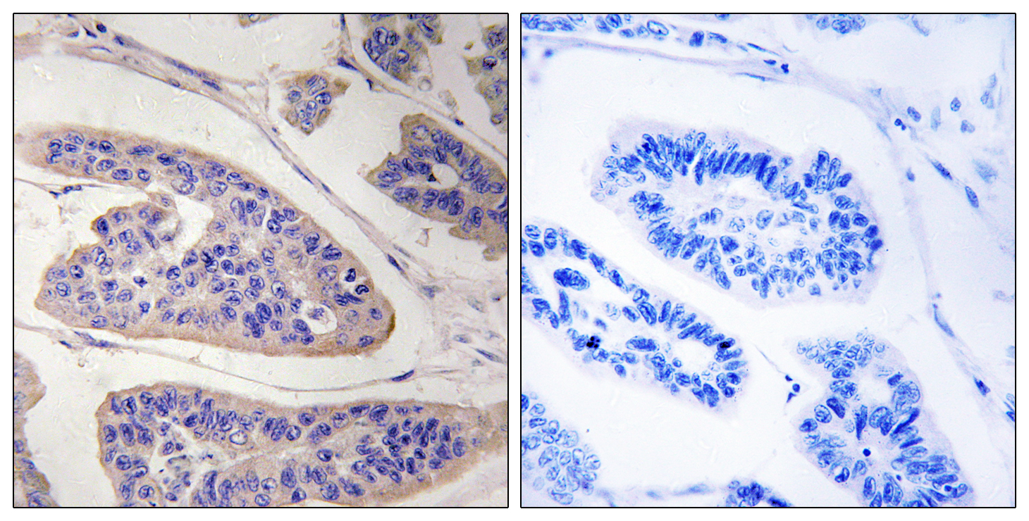
- Immunohistochemistry analysis of paraffin-embedded human breast carcinoma tissue, using ENAH Antibody. The picture on the right is blocked with the synthesized peptide.

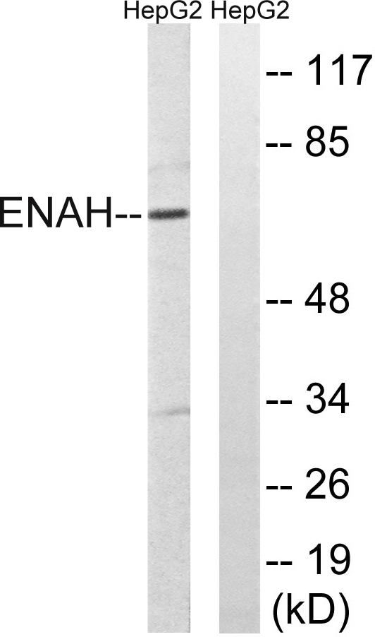
- Western blot analysis of lysates from HepG2 cells, using ENAH Antibody. The lane on the right is blocked with the synthesized peptide.


