Melan-A Polyclonal Antibody
- Catalog No.:YT2728
- Applications:WB;IHC;IF;ELISA
- Reactivity:Human;Rat;Mouse;
- Target:
- Melan-A
- Gene Name:
- MLANA
- Protein Name:
- Melanoma antigen recognized by T-cells 1
- Human Gene Id:
- 2315
- Human Swiss Prot No:
- Q16655
- Immunogen:
- The antiserum was produced against synthesized peptide derived from human MART-1. AA range:41-90
- Specificity:
- Melan-A Polyclonal Antibody detects endogenous levels of Melan-A protein.
- Formulation:
- Liquid in PBS containing 50% glycerol, 0.5% BSA and 0.02% sodium azide.
- Source:
- Polyclonal, Rabbit,IgG
- Dilution:
- WB 1:500 - 1:2000. IHC 1:100 - 1:300. ELISA: 1:40000.. IF 1:50-200
- Purification:
- The antibody was affinity-purified from rabbit antiserum by affinity-chromatography using epitope-specific immunogen.
- Concentration:
- 1 mg/ml
- Storage Stability:
- -15°C to -25°C/1 year(Do not lower than -25°C)
- Other Name:
- MLANA;MART1;Melanoma antigen recognized by T-cells 1;MART-1;Antigen LB39-AA;Antigen SK29-AA;Protein Melan-A
- Observed Band(KD):
- 15kD
- Background:
- tissue specificity:Expression is restricted to melanoma and melanocyte cell lines and retina.,
- Function:
- tissue specificity:Expression is restricted to melanoma and melanocyte cell lines and retina.,
- Subcellular Location:
- Endoplasmic reticulum membrane; Single-pass type III membrane protein. Golgi apparatus. Golgi apparatus, trans-Golgi network membrane. Melanosome. Also found in small vesicles and tubules dispersed over the entire cytoplasm. A small fraction of the protein is inserted into the membrane in an inverted orientation. Inversion of membrane topology results in the relocalization of the protein from a predominant Golgi/post-Golgi area to the endoplasmic reticulum. Melanoma cells expressing the protein with an inverted membrane topology are more effectively recognized by specific cytolytic T-lymphocytes than those expressing the protein in its native membrane orientation.
- Expression:
- Expression is restricted to melanoma and melanocyte cell lines and retina.
- June 19-2018
- WESTERN IMMUNOBLOTTING PROTOCOL
- June 19-2018
- IMMUNOHISTOCHEMISTRY-PARAFFIN PROTOCOL
- June 19-2018
- IMMUNOFLUORESCENCE PROTOCOL
- September 08-2020
- FLOW-CYTOMEYRT-PROTOCOL
- May 20-2022
- Cell-Based ELISA│解您多样本WB检测之困扰
- July 13-2018
- CELL-BASED-ELISA-PROTOCOL-FOR-ACETYL-PROTEIN
- July 13-2018
- CELL-BASED-ELISA-PROTOCOL-FOR-PHOSPHO-PROTEIN
- July 13-2018
- Antibody-FAQs
- Products Images
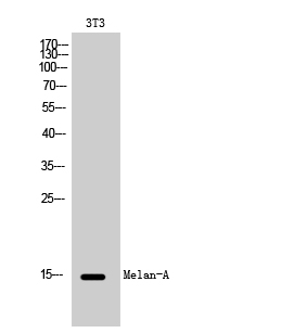
- Western Blot analysis of 3T3 cells using Melan-A Polyclonal Antibody
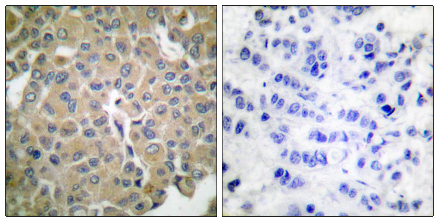
- Immunohistochemistry analysis of paraffin-embedded human breast carcinoma tissue, using MART-1 Antibody. The picture on the right is blocked with the synthesized peptide.
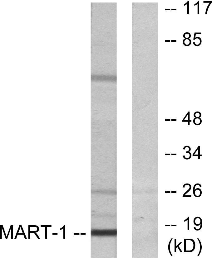
- Western blot analysis of lysates from NIH/3T3 cells, using MART-1 Antibody. The lane on the right is blocked with the synthesized peptide.


