Mat1 Polyclonal Antibody
- Catalog No.:YT2662
- Applications:WB;IHC;IF;ELISA
- Reactivity:Human;Mouse;Rat
- Target:
- Mat1
- Fields:
- >>Basal transcription factors;>>Nucleotide excision repair
- Gene Name:
- MNAT1
- Protein Name:
- CDK-activating kinase assembly factor MAT1
- Human Gene Id:
- 4331
- Human Swiss Prot No:
- P51948
- Mouse Gene Id:
- 17420
- Mouse Swiss Prot No:
- P51949
- Immunogen:
- The antiserum was produced against synthesized peptide derived from human MAT1. AA range:91-140
- Specificity:
- Mat1 Polyclonal Antibody detects endogenous levels of Mat1 protein.
- Formulation:
- Liquid in PBS containing 50% glycerol, 0.5% BSA and 0.02% sodium azide.
- Source:
- Polyclonal, Rabbit,IgG
- Dilution:
- WB 1:500 - 1:2000. IHC 1:100 - 1:300. ELISA: 1:20000.. IF 1:50-200
- Purification:
- The antibody was affinity-purified from rabbit antiserum by affinity-chromatography using epitope-specific immunogen.
- Concentration:
- 1 mg/ml
- Storage Stability:
- -15°C to -25°C/1 year(Do not lower than -25°C)
- Other Name:
- MNAT1;CAP35;MAT1;RNF66;CDK-activating kinase assembly factor MAT1;CDK7/cyclin-H assembly factor;Cyclin-G1-interacting protein;Menage a trois;RING finger protein 66;RING finger protein MAT1;p35;p36
- Observed Band(KD):
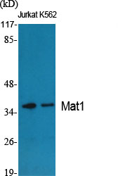
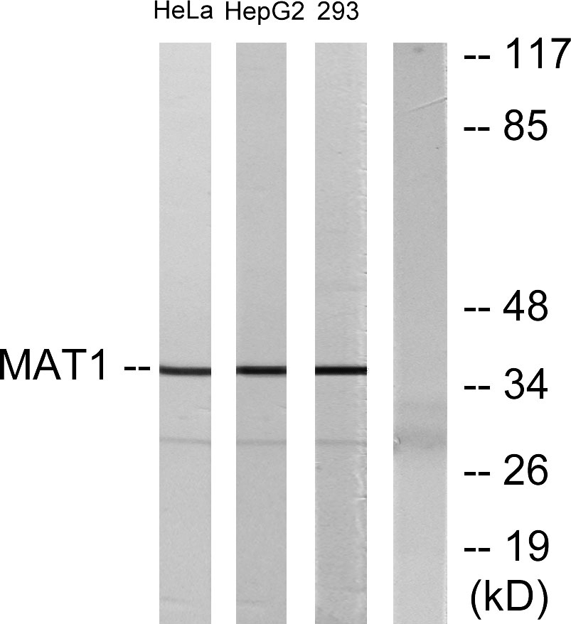
- 36kD
- Background:
- The protein encoded by this gene, along with cyclin H and CDK7, forms the CDK-activating kinase (CAK) enzymatic complex. This complex activates several cyclin-associated kinases and can also associate with TFIIH to activate transcription by RNA polymerase II. Two transcript variants encoding different isoforms have been found for this gene. [provided by RefSeq, Sep 2011],
- Function:
- function:Stabilizes the cyclin H-CDK7 complex to form a functional CDK-activating kinase (CAK) enzymatic complex. CAK activates the cyclin-associated kinases CDC2/CDK1, CDK2, CDK4 and CDK6 by threonine phosphorylation. CAK complexed to the core-TFIIH basal transcription factor activates RNA polymerase II by serine phosphorylation of the repetitive C-terminus domain (CTD) of its large subunit (POLR2A), allowing its escape from the promoter and elongation of the transcripts. Involved in cell cycle control and in RNA transcription by RNA polymerase II.,similarity:Contains 1 RING-type zinc finger.,similarity:Contains 1 UIM (ubiquitin-interacting motif) repeat.,subunit:Associates primarily with CDK7 and cyclin H to form the CAK complex. CAK can further associate with the core-TFIIH to form the TFIIH basal transcription factor.,tissue specificity:Highest levels in colon and testis. Moderate le
- Subcellular Location:
- Nucleus.
- Expression:
- Highest levels in colon and testis. Moderate levels are present thymus, prostate, ovary, and small intestine. The lowest levels are found in spleen and leukocytes.
- June 19-2018
- WESTERN IMMUNOBLOTTING PROTOCOL
- June 19-2018
- IMMUNOHISTOCHEMISTRY-PARAFFIN PROTOCOL
- June 19-2018
- IMMUNOFLUORESCENCE PROTOCOL
- September 08-2020
- FLOW-CYTOMEYRT-PROTOCOL
- May 20-2022
- Cell-Based ELISA│解您多样本WB检测之困扰
- July 13-2018
- CELL-BASED-ELISA-PROTOCOL-FOR-ACETYL-PROTEIN
- July 13-2018
- CELL-BASED-ELISA-PROTOCOL-FOR-PHOSPHO-PROTEIN
- July 13-2018
- Antibody-FAQs
- Products Images

- Western Blot analysis of various cells using Mat1 Polyclonal Antibody
.jpg)
- Western Blot analysis of 293 cells using Mat1 Polyclonal Antibody

- Western blot analysis of lysates from HeLa, HepG2, and 293 cells, using MAT1 Antibody. The lane on the right is blocked with the synthesized peptide.

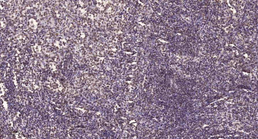
- Immunohistochemical analysis of paraffin-embedded human spleen. 1, Tris-EDTA,pH9.0 was used for antigen retrieval. 2 Antibody was diluted at 1:200(4° overnight.3,Secondary antibody was diluted at 1:200(room temperature, 45min).


