MARCH1 Polyclonal Antibody
- Catalog No.:YT2642
- Applications:IHC;IF;WB;ELISA
- Reactivity:Human;Mouse
- Target:
- MARCH1
- Gene Name:
- MARCH1
- Protein Name:
- E3 ubiquitin-protein ligase MARCH1
- Human Gene Id:
- 55016
- Human Swiss Prot No:
- Q8TCQ1
- Mouse Swiss Prot No:
- Q6NZQ8
- Immunogen:
- Synthesized peptide derived from the Internal region of human 40603.
- Specificity:
- MARCH1 Polyclonal Antibody detects endogenous levels of 40603 protein.
- Formulation:
- Liquid in PBS containing 50% glycerol, 0.5% BSA and 0.02% sodium azide.
- Source:
- Polyclonal, Rabbit,IgG
- Dilution:
- WB 1:500-2000 IHC 1:100 - 1:300. ELISA: 1:40000.. IF 1:50-200
- Purification:
- The antibody was affinity-purified from rabbit antiserum by affinity-chromatography using epitope-specific immunogen.
- Concentration:
- 1 mg/ml
- Storage Stability:
- -15°C to -25°C/1 year(Do not lower than -25°C)
- Other Name:
- MARCH1;RNF171;E3 ubiquitin-protein ligase MARCH1;Membrane-associated RING finger protein 1;Membrane-associated RING-CH protein I;MARCH-I;RING finger protein 171
- Observed Band(KD):
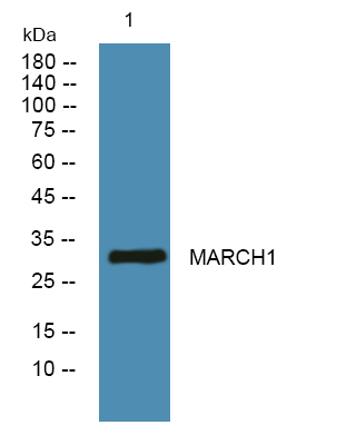
- 32kD
- Background:
- MARCH1 is a member of the MARCH family of membrane-bound E3 ubiquitin ligases (EC 6.3.2.19). MARCH proteins add ubiquitin (see MIM 191339) to target lysines in substrate proteins, thereby signaling their vesicular transport between membrane compartments. MARCH1 downregulates the surface expression of major histocompatibility complex (MHC) class II molecules (see MIM 142880) and other glycoproteins by directing them to the late endosomal/lysosomal compartment (Bartee et al., 2004 [PubMed 14722266]; Thibodeau et al., 2008 [PubMed 18389477]; De Gassart et al., 2008 [PubMed 18305173]).[supplied by OMIM, Mar 2010],
- Function:
- domain:The RING-CH-type zinc finger domain is required for E3 ligase activity.,function:E3 ubiquitin-protein ligase that may mediate ubiquitination of TFRC, CD86 and FAS, and promote their subsequent endocytosis and sorting to lysosomes via multivesicular bodies. E3 ubiquitin ligases accept ubiquitin from an E2 ubiquitin-conjugating enzyme in the form of a thioester and then directly transfer the ubiquitin to targeted substrates.,pathway:Protein modification; protein ubiquitination.,similarity:Contains 1 RING-CH-type zinc finger.,tissue specificity:Expressed in lymph nodes, spleen and lung.,
- Subcellular Location:
- Golgi apparatus, trans-Golgi network membrane ; Multi-pass membrane protein . Lysosome membrane ; Multi-pass membrane protein . Cytoplasmic vesicle membrane ; Multi-pass membrane protein . Late endosome membrane ; Multi-pass membrane protein . Early endosome membrane ; Multi-pass membrane protein . Cell membrane ; Multi-pass membrane protein .
- Expression:
- Expressed in antigen presenting cells, APCs, located in lymph nodes and spleen. Also expressed in lung. Expression is high in follicular B-cells, moderate in dendritic cells and low in splenic T-cells.
Myricetin Induces Autophagy and Cell Cycle Arrest of HCC by Inhibiting MARCH1-Regulated Stat3 and p38 MAPK Signaling Pathways. Frontiers in Pharmacology Front Pharmacol. 2021; 12: 709526 IHC Mouse 1:200 HepG2s-xenograft
Resveratrol inhibits the malignant progression of hepatocellular carcinoma via MARCH1-induced regulation of PTEN/AKT signaling. Aging-US Aging-Us. 2020 Jun 30; 12(12): 11717–11731 IHC Mouse 1:200 HepG2 cell-Xenograft
5‐FU inhibits migration and invasion of CRC cells through PI3K/AKT pathway regulated by MARCH1. CELL BIOLOGY INTERNATIONAL Cell Biol Int. 2021 Feb;45(2):368-381 WB Human Colorectal cancer(CRC ) tissues sw480s,DLD-1s,NCM460s
Wang, Nuan, et al. "MARCH1 Interacts with β-Catenin as a New Oncogene for Gastric Cancer." (2021).
5‐FU inhibits migration and invasion of CRC cells through PI3K/AKT pathway regulated by MARCH1. CELL BIOLOGY INTERNATIONAL Cell Biol Int. 2021 Feb;45(2):368-381 WB Human Colorectal cancer(CRC ) tissues sw480s,DLD-1s,NCM460s
MARCH1 negatively regulates TBK1-mTOR signaling pathway by ubiquitinating TBK1 BMC CANCER Li Xiao WB Human MCF7 cell,MFM223 cell,Sum159 cell,HEK293T cell
- June 19-2018
- WESTERN IMMUNOBLOTTING PROTOCOL
- June 19-2018
- IMMUNOHISTOCHEMISTRY-PARAFFIN PROTOCOL
- June 19-2018
- IMMUNOFLUORESCENCE PROTOCOL
- September 08-2020
- FLOW-CYTOMEYRT-PROTOCOL
- May 20-2022
- Cell-Based ELISA│解您多样本WB检测之困扰
- July 13-2018
- CELL-BASED-ELISA-PROTOCOL-FOR-ACETYL-PROTEIN
- July 13-2018
- CELL-BASED-ELISA-PROTOCOL-FOR-PHOSPHO-PROTEIN
- July 13-2018
- Antibody-FAQs
- Products Images

- Western blot analysis of lysates from DU145 cells, primary antibody was diluted at 1:1000, 4°over night

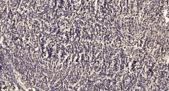
- Immunohistochemical analysis of paraffin-embedded human meningioma. 1, Antibody was diluted at 1:200(4° overnight). 2, Tris-EDTA,pH9.0 was used for antigen retrieval. 3,Secondary antibody was diluted at 1:200(room temperature, 45min).


