Laminin γ-1 Polyclonal Antibody
- Catalog No.:YT2531
- Applications:WB;IHC;IF;ELISA
- Reactivity:Human;Mouse;Rat;Monkey;Cat
- Target:
- Laminin γ-1
- Fields:
- >>PI3K-Akt signaling pathway;>>Focal adhesion;>>ECM-receptor interaction;>>Prion disease;>>Toxoplasmosis;>>Amoebiasis;>>Human papillomavirus infection;>>Pathways in cancer;>>Small cell lung cancer
- Gene Name:
- LAMC1
- Protein Name:
- Laminin subunit gamma-1
- Human Gene Id:
- 3915
- Human Swiss Prot No:
- P11047
- Mouse Swiss Prot No:
- P02468
- Immunogen:
- The antiserum was produced against synthesized peptide derived from human LAMC1. AA range:1451-1500
- Specificity:
- Laminin γ-1 Polyclonal Antibody detects endogenous levels of Laminin γ-1 protein.
- Formulation:
- Liquid in PBS containing 50% glycerol, 0.5% BSA and 0.02% sodium azide.
- Source:
- Polyclonal, Rabbit,IgG
- Dilution:
- WB 1:500 - 1:2000. IHC 1:100 - 1:300. IF 1:200 - 1:1000. ELISA: 1:40000. Not yet tested in other applications.
- Purification:
- The antibody was affinity-purified from rabbit antiserum by affinity-chromatography using epitope-specific immunogen.
- Concentration:
- 1 mg/ml
- Storage Stability:
- -15°C to -25°C/1 year(Do not lower than -25°C)
- Other Name:
- LAMC1;LAMB2;Laminin subunit gamma-1;Laminin B2 chain;Laminin-1 subunit gamma;Laminin-10 subunit gamma;Laminin-11 subunit gamma;Laminin-2 subunit gamma;Laminin-3 subunit gamma;Laminin-4 subunit gamma;Laminin-6 subunit gamma;Lamini
- Observed Band(KD):
- 178kD
- Background:
- Laminins, a family of extracellular matrix glycoproteins, are the major noncollagenous constituent of basement membranes. They have been implicated in a wide variety of biological processes including cell adhesion, differentiation, migration, signaling, neurite outgrowth and metastasis. Laminins, composed of 3 non identical chains: laminin alpha, beta and gamma (formerly A, B1, and B2, respectively), have a cruciform structure consisting of 3 short arms, each formed by a different chain, and a long arm composed of all 3 chains. Each laminin chain is a multidomain protein encoded by a distinct gene. Several isoforms of each chain have been described. Different alpha, beta and gamma chain isomers combine to give rise to different heterotrimeric laminin isoforms which are designated by Arabic numerals in the order of their discovery, i.e. alpha1beta1gamma1 heterotrimer is laminin 1. The biological func
- Function:
- domain:Domains VI and IV are globular.,domain:The alpha-helical domains I and II are thought to interact with other laminin chains to form a coiled coil structure.,function:Binding to cells via a high affinity receptor, laminin is thought to mediate the attachment, migration and organization of cells into tissues during embryonic development by interacting with other extracellular matrix components.,similarity:Contains 1 laminin IV type A domain.,similarity:Contains 1 laminin N-terminal domain.,similarity:Contains 11 laminin EGF-like domains.,subunit:Laminin is a complex glycoprotein, consisting of three different polypeptide chains (alpha, beta, gamma), which are bound to each other by disulfide bonds into a cross-shaped molecule comprising one long and three short arms with globules at each end. Gamma-1 is a subunit of laminin-1 (EHS laminin), laminin-2 (merosin), laminin-3 (S-laminin)
- Subcellular Location:
- Secreted, extracellular space, extracellular matrix, basement membrane.
- Expression:
- Found in the basement membranes (major component).
Profiling of Host Cell Response to Successive Canine Parvovirus Infection Based on Kinetic Proteomic Change Identification. Scientific Reports 2016 Jul 13 WB Cat F81 cell
- June 19-2018
- WESTERN IMMUNOBLOTTING PROTOCOL
- June 19-2018
- IMMUNOHISTOCHEMISTRY-PARAFFIN PROTOCOL
- June 19-2018
- IMMUNOFLUORESCENCE PROTOCOL
- September 08-2020
- FLOW-CYTOMEYRT-PROTOCOL
- May 20-2022
- Cell-Based ELISA│解您多样本WB检测之困扰
- July 13-2018
- CELL-BASED-ELISA-PROTOCOL-FOR-ACETYL-PROTEIN
- July 13-2018
- CELL-BASED-ELISA-PROTOCOL-FOR-PHOSPHO-PROTEIN
- July 13-2018
- Antibody-FAQs
- Products Images
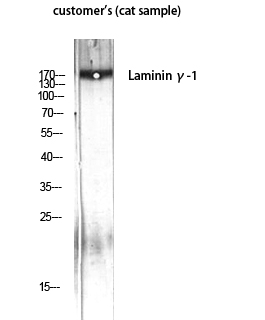
- Western Blot analysis of customer's (cat sample) using Laminin γ-1 Polyclonal Antibody diluted at 1:1000

- Western blot analysis of lysates from HUVEC cells, using LAMC1 Antibody. The lane on the right is blocked with the synthesized peptide.
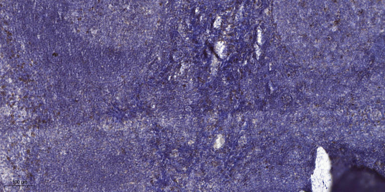
- Immunohistochemical analysis of paraffin-embedded human tonsil. 1, Antibody was diluted at 1:200(4° overnight). 2, Tris-EDTA,pH9.0 was used for antigen retrieval. 3,Secondary antibody was diluted at 1:200(room temperature, 30min).


