Lamin A/C Polyclonal Antibody
- Catalog No.:YT2520
- Applications:WB;IHC;IF;ELISA
- Reactivity:Human;Mouse;Rat
- Target:
- Lamin A/C
- Fields:
- >>Apoptosis;>>Hypertrophic cardiomyopathy;>>Arrhythmogenic right ventricular cardiomyopathy;>>Dilated cardiomyopathy
- Gene Name:
- LMNA
- Protein Name:
- Prelamin-A/C
- Human Gene Id:
- 4000
- Human Swiss Prot No:
- P02545
- Mouse Gene Id:
- 16905
- Mouse Swiss Prot No:
- P48678
- Rat Gene Id:
- 60374
- Rat Swiss Prot No:
- P48679
- Immunogen:
- The antiserum was produced against synthesized peptide derived from human Lamin A/C. AA range:361-410
- Specificity:
- Lamin A/C Polyclonal Antibody detects endogenous levels of Lamin A/C protein.
- Formulation:
- Liquid in PBS containing 50% glycerol, 0.5% BSA and 0.02% sodium azide.
- Source:
- Polyclonal, Rabbit,IgG
- Dilution:
- WB 1:500 - 1:2000. IHC 1:100 - 1:300. IF 1:200 - 1:1000. ELISA: 1:20000. Not yet tested in other applications.
- Purification:
- The antibody was affinity-purified from rabbit antiserum by affinity-chromatography using epitope-specific immunogen.
- Concentration:
- 1 mg/ml
- Storage Stability:
- -15°C to -25°C/1 year(Do not lower than -25°C)
- Other Name:
- LMNA;LMN1;Prelamin-A/C
- Observed Band(KD):
- 74kD,65kD
- Background:
- lamin A/C(LMNA) Homo sapiens The nuclear lamina consists of a two-dimensional matrix of proteins located next to the inner nuclear membrane. The lamin family of proteins make up the matrix and are highly conserved in evolution. During mitosis, the lamina matrix is reversibly disassembled as the lamin proteins are phosphorylated. Lamin proteins are thought to be involved in nuclear stability, chromatin structure and gene expression. Vertebrate lamins consist of two types, A and B. Alternative splicing results in multiple transcript variants. Mutations in this gene lead to several diseases: Emery-Dreifuss muscular dystrophy, familial partial lipodystrophy, limb girdle muscular dystrophy, dilated cardiomyopathy, Charcot-Marie-Tooth disease, and Hutchinson-Gilford progeria syndrome. [provided by RefSeq, Apr 2012],
- Function:
- disease:Defects in LMNA are a cause of Emery-Dreifuss muscular dystrophy type 2 (EDMD2) [MIM:181350]. EDMD2 is an autosomal dominant disorder characterized by slowly progressive muscle wasting and weakness, early contractures of the elbows Achilles tendons and spine, and cardiomyopathy associated with cardiac conduction defects.,disease:Defects in LMNA are a cause of Emery-Dreifuss muscular dystrophy type 3 (EDMD3) [MIM:604929]. EDMD3 is an autosomal recessive disorder characterized by early contractures, muscle wasting and weakness and cardiomyopathy.,disease:Defects in LMNA are a cause of familial partial lipodystrophy type 2 (FPLD2) [MIM:151660]; also known as familial partial lipodystrophy Dunnigan type. FPLD2 is an autosomal dominant disorder characterized by marked loss of subcutaneous adipose tissue from the extremities and trunk but by excess fat deposition in the head and neck.
- Subcellular Location:
- Nucleus . Nucleus envelope . Nucleus lamina. Nucleus, nucleoplasm. Nucleus matrix . Farnesylation of prelamin-A/C facilitates nuclear envelope targeting and subsequent cleavage by ZMPSTE24/FACE1 to remove the farnesyl group produces mature lamin-A/C, which can then be inserted into the nuclear lamina. EMD is required for proper localization of non-farnesylated prelamin-A/C.; [Isoform C]: Nucleus speckle .
- Expression:
- In the arteries, prelamin-A/C accumulation is not observed in young healthy vessels but is prevalent in medial vascular smooth muscle cells (VSMCs) from aged individuals and in atherosclerotic lesions, where it often colocalizes with senescent and degenerate VSMCs. Prelamin-A/C expression increases with age and disease. In normal aging, the accumulation of prelamin-A/C is caused in part by the down-regulation of ZMPSTE24/FACE1 in response to oxidative stress.
A Novel Function of F-Box Protein FBXO17 in Negative Regulation of Type I IFN Signaling by Recruiting PP2A for IFN Regulatory Factor 3 Deactivation. JOURNAL OF IMMUNOLOGY J Immunol. 2017 Jan;198(2):808-819 WB Human HEK293T cell
Ganglioside GD1a suppresses LPS-induced pro-inflammatory cytokines in RAW264.7 macrophages by reducing MAPKs and NF-κB signaling pathways through TLR4. INTERNATIONAL IMMUNOPHARMACOLOGY Int Immunopharmacol. 2015 Sep;28:136 WB Mouse RAW264.7 Cell
- June 19-2018
- WESTERN IMMUNOBLOTTING PROTOCOL
- June 19-2018
- IMMUNOHISTOCHEMISTRY-PARAFFIN PROTOCOL
- June 19-2018
- IMMUNOFLUORESCENCE PROTOCOL
- September 08-2020
- FLOW-CYTOMEYRT-PROTOCOL
- May 20-2022
- Cell-Based ELISA│解您多样本WB检测之困扰
- July 13-2018
- CELL-BASED-ELISA-PROTOCOL-FOR-ACETYL-PROTEIN
- July 13-2018
- CELL-BASED-ELISA-PROTOCOL-FOR-PHOSPHO-PROTEIN
- July 13-2018
- Antibody-FAQs
- Products Images
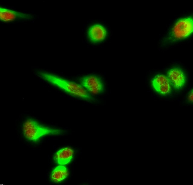
- Immunofluorescence analysis of Hela cell. 1,Lamin A/C Polyclonal Antibody(red) was diluted at 1:200(4° overnight). Galectin-3 Monoclonal Antibody(6G2)(green) was diluted at 1:200(4° overnight). 2, Goat Anti Rabbit Alexa Fluor 594 Catalog:RS3611 was diluted at 1:1000(room temperature, 50min). Goat Anti Mouse Alexa Fluor 488 Catalog:RS3208 was diluted at 1:1000(room temperature, 50min).
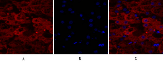
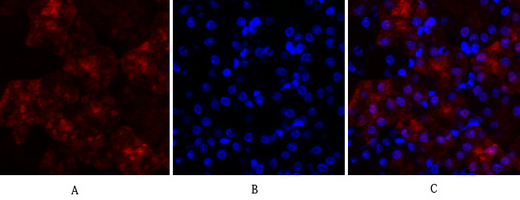
- Immunofluorescence analysis of human-liver tissue. 1,Lamin A/C Polyclonal Antibody(red) was diluted at 1:200(4°C,overnight). 2, Cy3 labled Secondary antibody was diluted at 1:300(room temperature, 50min).3, Picture B: DAPI(blue) 10min. Picture A:Target. Picture B: DAPI. Picture C: merge of A+B
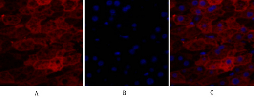
- Immunofluorescence analysis of human-liver tissue. 1,Lamin A/C Polyclonal Antibody(red) was diluted at 1:200(4°C,overnight). 2, Cy3 labled Secondary antibody was diluted at 1:300(room temperature, 50min).3, Picture B: DAPI(blue) 10min. Picture A:Target. Picture B: DAPI. Picture C: merge of A+B
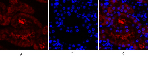
- Immunofluorescence analysis of rat-kidney tissue. 1,Lamin A/C Polyclonal Antibody(red) was diluted at 1:200(4°C,overnight). 2, Cy3 labled Secondary antibody was diluted at 1:300(room temperature, 50min).3, Picture B: DAPI(blue) 10min. Picture A:Target. Picture B: DAPI. Picture C: merge of A+B
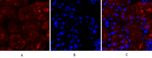
- Immunofluorescence analysis of rat-kidney tissue. 1,Lamin A/C Polyclonal Antibody(red) was diluted at 1:200(4°C,overnight). 2, Cy3 labled Secondary antibody was diluted at 1:300(room temperature, 50min).3, Picture B: DAPI(blue) 10min. Picture A:Target. Picture B: DAPI. Picture C: merge of A+B
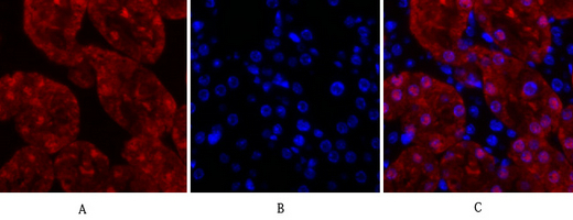
- Immunofluorescence analysis of mouse-kidney tissue. 1,Lamin A/C Polyclonal Antibody(red) was diluted at 1:200(4°C,overnight). 2, Cy3 labled Secondary antibody was diluted at 1:300(room temperature, 50min).3, Picture B: DAPI(blue) 10min. Picture A:Target. Picture B: DAPI. Picture C: merge of A+B
- Immunofluorescence analysis of mouse-kidney tissue. 1,Lamin A/C Polyclonal Antibody(red) was diluted at 1:200(4°C,overnight). 2, Cy3 labled Secondary antibody was diluted at 1:300(room temperature, 50min).3, Picture B: DAPI(blue) 10min. Picture A:Target. Picture B: DAPI. Picture C: merge of A+B
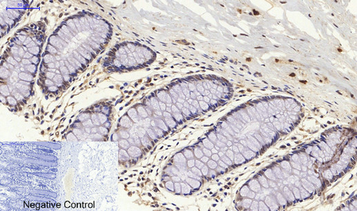
- Immunohistochemical analysis of paraffin-embedded Human-colon tissue. 1,Lamin A/C Polyclonal Antibody was diluted at 1:200(4°C,overnight). 2, Sodium citrate pH 6.0 was used for antibody retrieval(>98°C,20min). 3,Secondary antibody was diluted at 1:200(room tempeRature, 30min). Negative control was used by secondary antibody only.
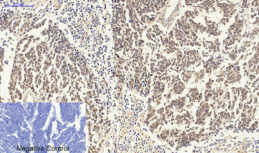
- Immunohistochemical analysis of paraffin-embedded Human-lung-cancer tissue. 1,Lamin A/C Polyclonal Antibody was diluted at 1:200(4°C,overnight). 2, Sodium citrate pH 6.0 was used for antibody retrieval(>98°C,20min). 3,Secondary antibody was diluted at 1:200(room tempeRature, 30min). Negative control was used by secondary antibody only.

- Immunohistochemical analysis of paraffin-embedded Human-stomach-cancer tissue. 1,Lamin A/C Polyclonal Antibody was diluted at 1:200(4°C,overnight). 2, Sodium citrate pH 6.0 was used for antibody retrieval(>98°C,20min). 3,Secondary antibody was diluted at 1:200(room tempeRature, 30min). Negative control was used by secondary antibody only.
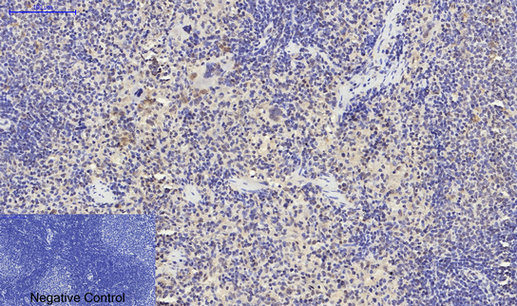
- Immunohistochemical analysis of paraffin-embedded Rat-spleen tissue. 1,Lamin A/C Polyclonal Antibody was diluted at 1:200(4°C,overnight). 2, Sodium citrate pH 6.0 was used for antibody retrieval(>98°C,20min). 3,Secondary antibody was diluted at 1:200(room tempeRature, 30min). Negative control was used by secondary antibody only.
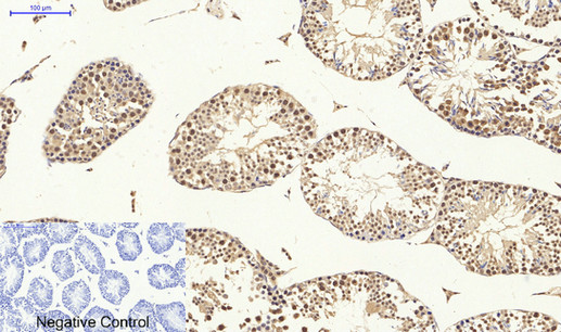
- Immunohistochemical analysis of paraffin-embedded Mouse-testis tissue. 1,Lamin A/C Polyclonal Antibody was diluted at 1:200(4°C,overnight). 2, Sodium citrate pH 6.0 was used for antibody retrieval(>98°C,20min). 3,Secondary antibody was diluted at 1:200(room tempeRature, 30min). Negative control was used by secondary antibody only.
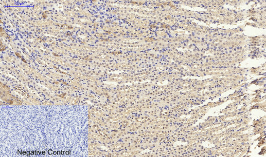
- Immunohistochemical analysis of paraffin-embedded Mouse-kidney tissue. 1,Lamin A/C Polyclonal Antibody was diluted at 1:200(4°C,overnight). 2, Sodium citrate pH 6.0 was used for antibody retrieval(>98°C,20min). 3,Secondary antibody was diluted at 1:200(room tempeRature, 30min). Negative control was used by secondary antibody only.
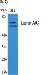
- Western Blot analysis of various cells using Lamin A/C Polyclonal Antibody diluted at 1:2000
.jpg)
- Western Blot analysis of HepG2 cells using Lamin A/C Polyclonal Antibody diluted at 1:2000
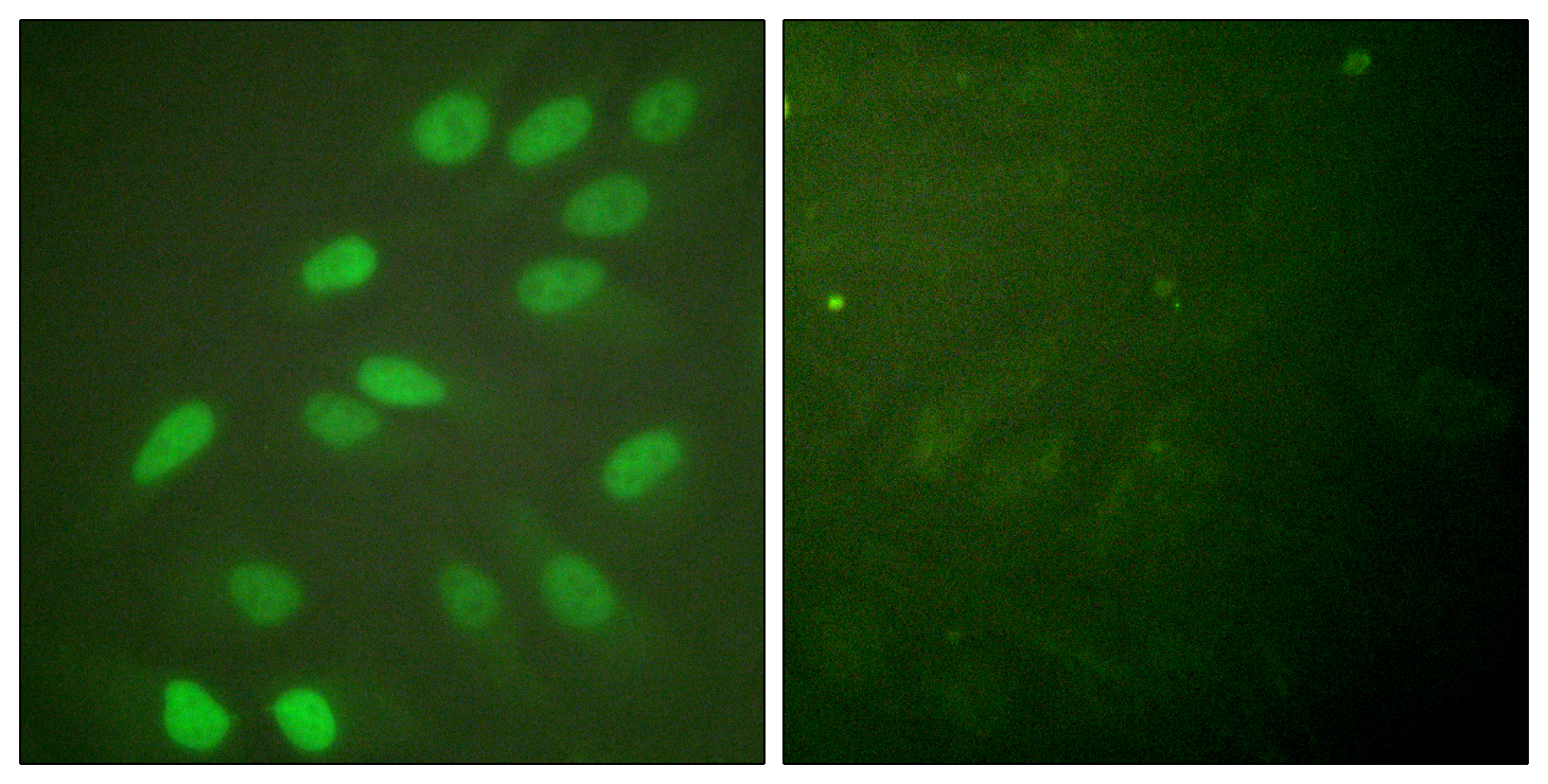
- Immunofluorescence analysis of HeLa cells, using Lamin A/C Antibody. The picture on the right is blocked with the synthesized peptide.
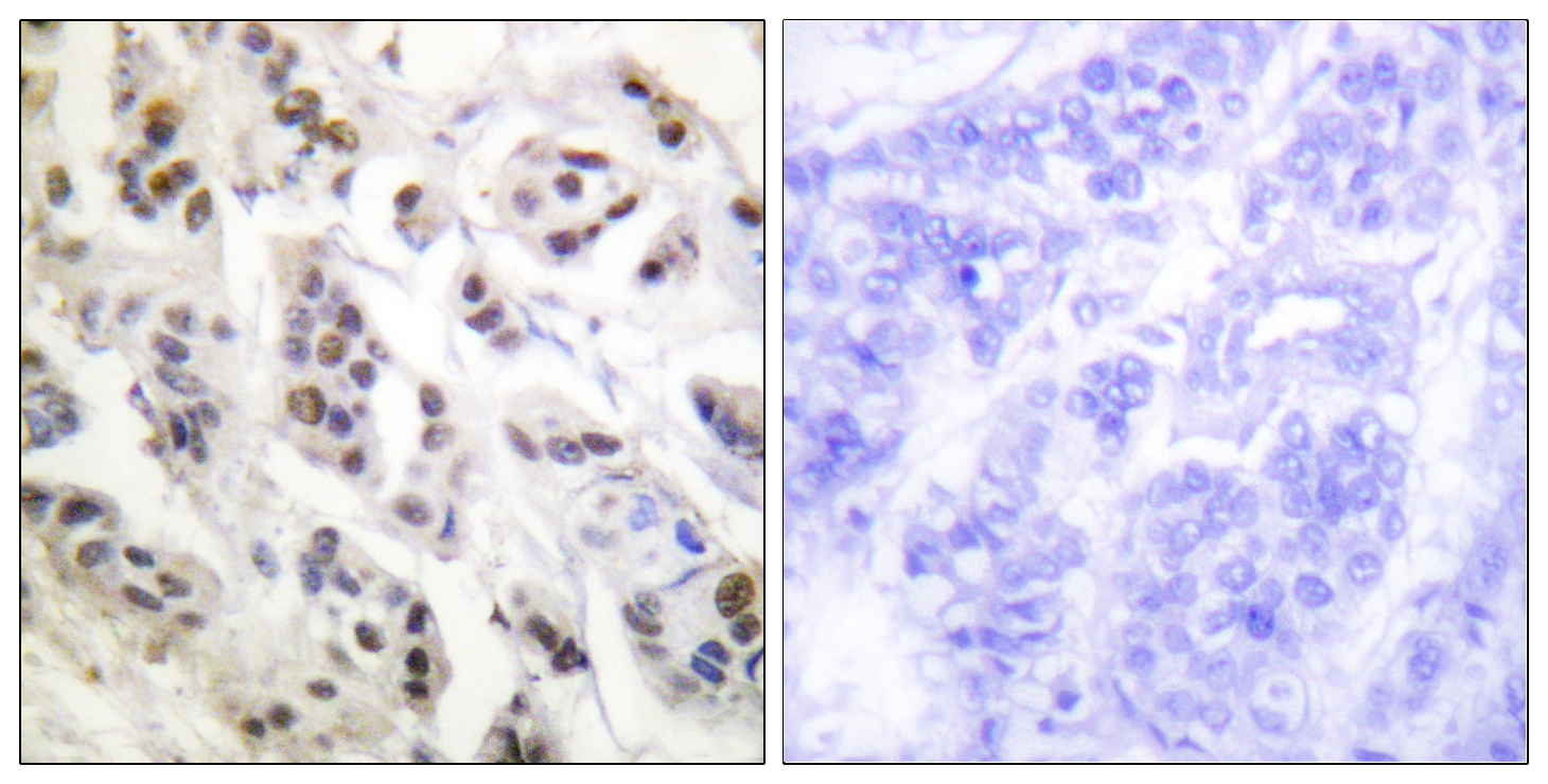
- Immunohistochemistry analysis of paraffin-embedded human breast carcinoma tissue, using Lamin A/C Antibody. The picture on the right is blocked with the synthesized peptide.
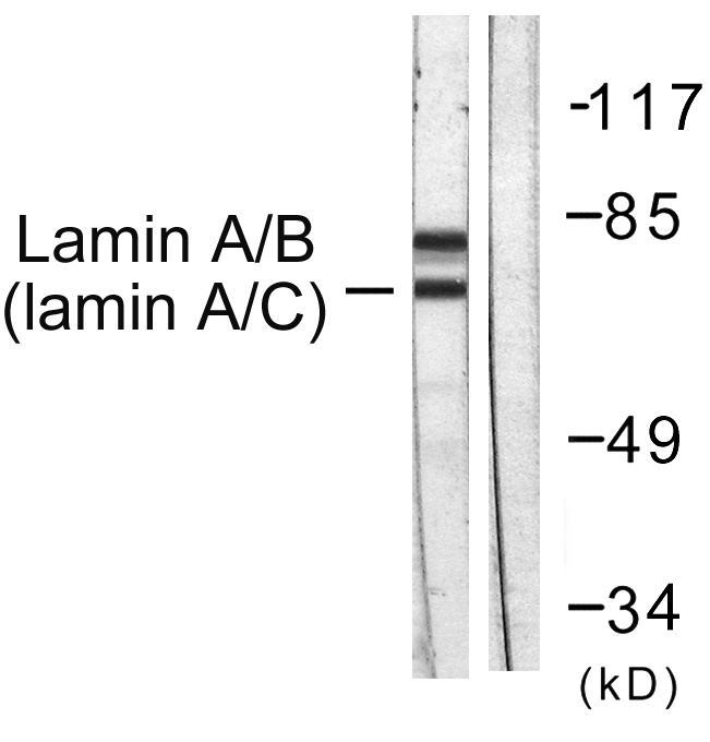
- Western blot analysis of lysates from HeLa cells, using Lamin A/C Antibody. The lane on the right is blocked with the synthesized peptide.


