KV8.2 Polyclonal Antibody
- Catalog No.:YT2515
- Applications:WB;IHC
- Reactivity:Human;Mouse
- Target:
- KV8.2
- Gene Name:
- KCNV2
- Protein Name:
- Potassium voltage-gated channel subfamily V member 2
- Human Gene Id:
- 169522
- Human Swiss Prot No:
- Q8TDN2
- Mouse Gene Id:
- 240595
- Mouse Swiss Prot No:
- Q8CFS6
- Immunogen:
- The antiserum was produced against synthesized peptide derived from human KCNV2. AA range:187-236
- Specificity:
- KV8.2 Polyclonal Antibody detects endogenous levels of KV8.2 protein.
- Formulation:
- Liquid in PBS containing 50% glycerol, 0.5% BSA and 0.02% sodium azide.
- Source:
- Polyclonal, Rabbit,IgG
- Dilution:
- WB 1:500-2000;IHC 1:50-300
- Purification:
- The antibody was affinity-purified from rabbit antiserum by affinity-chromatography using epitope-specific immunogen.
- Concentration:
- 1 mg/ml
- Storage Stability:
- -15°C to -25°C/1 year(Do not lower than -25°C)
- Other Name:
- KCNV2;Potassium voltage-gated channel subfamily V member 2;Voltage-gated potassium channel subunit Kv8.2
- Observed Band(KD):
- 62kD
- Background:
- Voltage-gated potassium (Kv) channels represent the most complex class of voltage-gated ion channels from both functional and structural standpoints. Their diverse functions include regulating neurotransmitter release, heart rate, insulin secretion, neuronal excitability, epithelial electrolyte transport, smooth muscle contraction, and cell volume. This gene encodes a member of the potassium voltage-gated channel subfamily V. This member is identified as a 'silent subunit', and it does not form homomultimers, but forms heteromultimers with several other subfamily members. Through obligatory heteromerization, it exerts a function-altering effect on other potassium channel subunits. This protein is strongly expressed in pancreas and has a weaker expression in several other tissues. [provided by RefSeq, Jul 2008],
- Function:
- disease:Defects in KCNV2 are the cause of cone dystrophy retinal type 3B (RCD3B) [MIM:610356]; also called cone dystrophy with night blindness and supernormal rod responses KCNV2-related. RCD3B is a rare form of cone dystrophy associated with supernormal rod responses. The disorder is characterized by reduced visual acuity, photoaversion, night blindness, and abnormal color vision. At an early age, the retina shows subtle depigmentation at the macula and, later, more obvious areas of atrophy.,domain:The segment S4 is probably the voltage-sensor and is characterized by a series of positively charged amino acids at every third position.,function:Potassium channel subunit. Modulates channel activity by shifting the threshold and the half-maximal activation to more negative values.,similarity:Belongs to the potassium channel family. V subfamily.,subcellular location:Has to be associated with
- Subcellular Location:
- Cell membrane; Multi-pass membrane protein. Has to be associated with KCNB1 or possibly another partner to get inserted in the plasma membrane. Remains intracellular in the absence of KCNB1.
- Expression:
- Detected in lung, liver, kidney, pancreas, spleen, thymus, prostate, testis, ovary and colon.
- June 19-2018
- WESTERN IMMUNOBLOTTING PROTOCOL
- June 19-2018
- IMMUNOHISTOCHEMISTRY-PARAFFIN PROTOCOL
- June 19-2018
- IMMUNOFLUORESCENCE PROTOCOL
- September 08-2020
- FLOW-CYTOMEYRT-PROTOCOL
- May 20-2022
- Cell-Based ELISA│解您多样本WB检测之困扰
- July 13-2018
- CELL-BASED-ELISA-PROTOCOL-FOR-ACETYL-PROTEIN
- July 13-2018
- CELL-BASED-ELISA-PROTOCOL-FOR-PHOSPHO-PROTEIN
- July 13-2018
- Antibody-FAQs
- Products Images

- Western Blot analysis of MCF-7 cells using KV8.2 Polyclonal Antibody
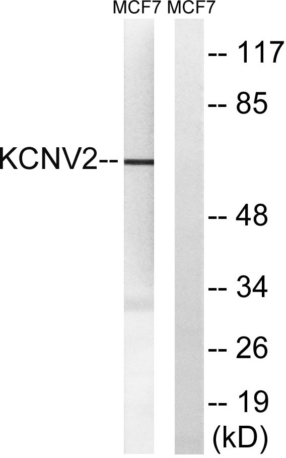
- Western blot analysis of lysates from MCF-7 cells, using KCNV2 Antibody. The lane on the right is blocked with the synthesized peptide.
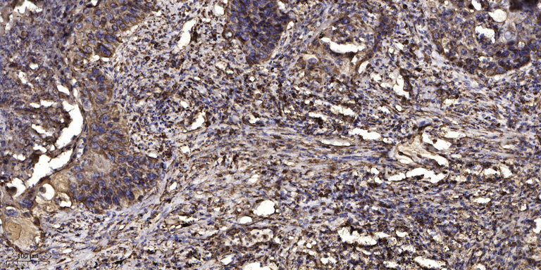
- Immunohistochemical analysis of paraffin-embedded human Squamous cell carcinoma of lung. 1, Antibody was diluted at 1:200(4° overnight). 2, Tris-EDTA,pH9.0 was used for antigen retrieval. 3,Secondary antibody was diluted at 1:200(room temperature, 45min).


