KV1.1 Polyclonal Antibody
- Catalog No.:YT2505
- Applications:WB;ELISA;IHC
- Reactivity:Human;Mouse;Rat
- Target:
- KV1.1
- Gene Name:
- KCNA1
- Protein Name:
- Potassium voltage-gated channel subfamily A member 1
- Human Gene Id:
- 3736
- Human Swiss Prot No:
- Q09470
- Mouse Gene Id:
- 16485
- Mouse Swiss Prot No:
- P16388
- Rat Gene Id:
- 24520
- Rat Swiss Prot No:
- P10499
- Immunogen:
- The antiserum was produced against synthesized peptide derived from human KCNA1. AA range:256-305
- Specificity:
- KV1.1 Polyclonal Antibody detects endogenous levels of KV1.1 protein.
- Formulation:
- Liquid in PBS containing 50% glycerol, 0.5% BSA and 0.02% sodium azide.
- Source:
- Polyclonal, Rabbit,IgG
- Dilution:
- WB 1:500-2000;IHC 1:50-300; ELISA 2000-20000
- Purification:
- The antibody was affinity-purified from rabbit antiserum by affinity-chromatography using epitope-specific immunogen.
- Concentration:
- 1 mg/ml
- Storage Stability:
- -15°C to -25°C/1 year(Do not lower than -25°C)
- Other Name:
- KCNA1;Potassium voltage-gated channel subfamily A member 1;Voltage-gated K(+) channel HuKI;Voltage-gated potassium channel HBK1;Voltage-gated potassium channel subunit Kv1.1
- Observed Band(KD):
- 57kD
- Background:
- This gene encodes a voltage-gated delayed potassium channel that is phylogenetically related to the Drosophila Shaker channel. The encoded protein has six putative transmembrane segments (S1-S6), and the loop between S5 and S6 forms the pore and contains the conserved selectivity filter motif (GYGD). The functional channel is a homotetramer. The N-terminus of the channel is associated with beta subunits that can modify the inactivation properties of the channel as well as affect expression levels. The C-terminus of the channel is complexed to a PDZ domain protein that is responsible for channel targeting. Mutations in this gene have been associated with myokymia with periodic ataxia (AEMK). [provided by RefSeq, Jul 2008],
- Function:
- disease:Defects in KCNA1 are the cause of episodic ataxia type 1 (EA1) [MIM:160120]; also known as paroxysmal or episodic ataxia with myokymia (EAM) or paroxysmal ataxia with neuromyotonia. EA1 is an autosomal dominant disorder characterized by brief episodes of ataxia and dysarthria. Neurological examination during and between the attacks demonstrates spontaneous, repetitive discharges in the distal musculature (myokymia) that arise from peripheral nerve. Nystagmus is absent.,disease:Defects in KCNA1 are the cause of myokymia isolated type 1 (MK1) [MIM:160120]. Myokymia is a condition characterized by spontaneous involuntary contraction of muscle fiber groups that can be observed as vermiform movement of the overlying skin. Electromyography typically shows continuous motor unit activity with spontaneous oligo- and multiplet-discharges of high intraburst frequency (myokymic discharges).
- Subcellular Location:
- Cell membrane ; Multi-pass membrane protein . Membrane . Cell projection, axon . Cytoplasmic vesicle . Perikaryon . Endoplasmic reticulum . Cell projection, dendrite . Cell junction . Cell junction, synapse . Cell junction, synapse, presynaptic cell membrane . Cell junction, synapse, presynapse . Homotetrameric KCNA1 is primarily located in the endoplasmic reticulum. Interaction with KCNA2 and KCNAB2 or with KCNA4 and KCNAB2 promotes expression at the cell membrane (By similarity). .
- Expression:
- Detected adjacent to nodes of Ranvier in juxtaparanodal zones in spinal cord nerve fibers, but also in paranodal regions in some myelinated spinal cord axons (at protein level) (PubMed:11086297). Detected in the islet of Langerhans (PubMed:21483673).
- June 19-2018
- WESTERN IMMUNOBLOTTING PROTOCOL
- June 19-2018
- IMMUNOHISTOCHEMISTRY-PARAFFIN PROTOCOL
- June 19-2018
- IMMUNOFLUORESCENCE PROTOCOL
- September 08-2020
- FLOW-CYTOMEYRT-PROTOCOL
- May 20-2022
- Cell-Based ELISA│解您多样本WB检测之困扰
- July 13-2018
- CELL-BASED-ELISA-PROTOCOL-FOR-ACETYL-PROTEIN
- July 13-2018
- CELL-BASED-ELISA-PROTOCOL-FOR-PHOSPHO-PROTEIN
- July 13-2018
- Antibody-FAQs
- Products Images
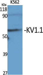
- Western Blot analysis of various cells using KV1.1 Polyclonal Antibody diluted at 1:2000
.jpg)
- Western Blot analysis of HeLa cells using KV1.1 Polyclonal Antibody diluted at 1:2000
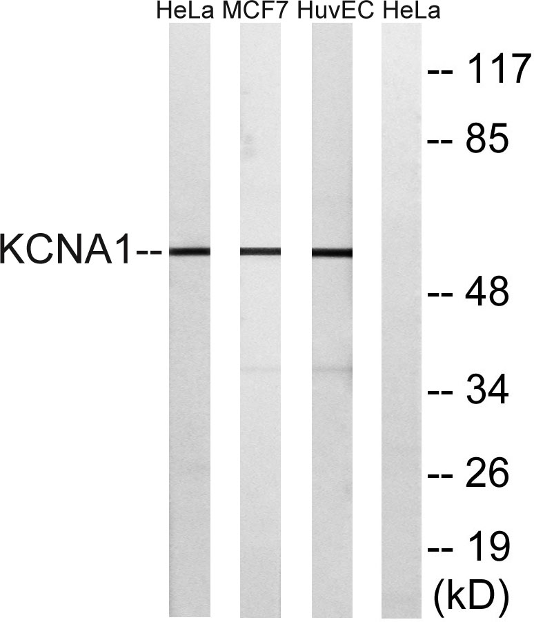
- Western blot analysis of lysates from HUVEC, MCF-7, and HeLa cells, using KCNA1 Antibody. The lane on the right is blocked with the synthesized peptide.
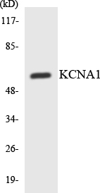
- Western blot analysis of the lysates from HepG2 cells using KCNA1 antibody.
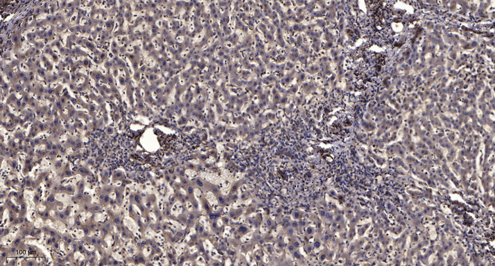
- Immunohistochemical analysis of paraffin-embedded human liver cancer. 1, Antibody was diluted at 1:200(4° overnight). 2, Tris-EDTA,pH9.0 was used for antigen retrieval. 3,Secondary antibody was diluted at 1:200(room temperature, 45min).


