JIP-2 Polyclonal Antibody
- Catalog No.:YT2435
- Applications:WB;IHC;IF;ELISA
- Reactivity:Human;Mouse;Rat
- Target:
- JIP-2
- Fields:
- >>MAPK signaling pathway
- Gene Name:
- MAPK8IP2
- Protein Name:
- C-Jun-amino-terminal kinase-interacting protein 2
- Human Gene Id:
- 23542
- Human Swiss Prot No:
- Q13387
- Mouse Gene Id:
- 60597
- Mouse Swiss Prot No:
- Q9ERE9
- Immunogen:
- The antiserum was produced against synthesized peptide derived from human JIP2. AA range:581-630
- Specificity:
- JIP-2 Polyclonal Antibody detects endogenous levels of JIP-2 protein.
- Formulation:
- Liquid in PBS containing 50% glycerol, 0.5% BSA and 0.02% sodium azide.
- Source:
- Polyclonal, Rabbit,IgG
- Dilution:
- WB 1:500 - 1:2000. IHC 1:100 - 1:300. IF 1:200 - 1:1000. ELISA: 1:20000. Not yet tested in other applications.
- Purification:
- The antibody was affinity-purified from rabbit antiserum by affinity-chromatography using epitope-specific immunogen.
- Concentration:
- 1 mg/ml
- Storage Stability:
- -15°C to -25°C/1 year(Do not lower than -25°C)
- Other Name:
- MAPK8IP2;IB2;JIP2;PRKM8IPL;C-Jun-amino-terminal kinase-interacting protein 2;JIP-2;JNK-interacting protein 2;Islet-brain-2;IB-2;JNK MAP kinase scaffold protein 2;Mitogen-activated protein kinase 8-interacting protein 2
- Observed Band(KD):
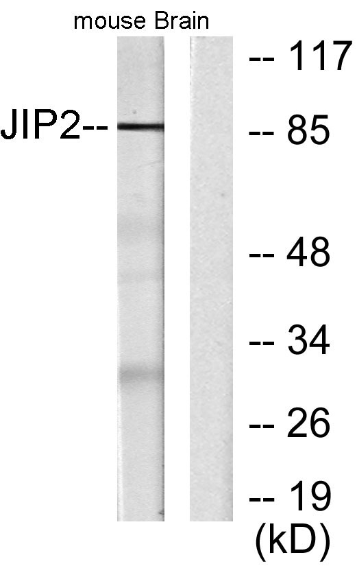
- 87kD
- Background:
- The protein encoded by this gene is closely related to MAPK8IP1/IB1/JIP-1, a scaffold protein that is involved in the c-Jun amino-terminal kinase signaling pathway. This protein is expressed in brain and pancreatic cells. It has been shown to interact with, and regulate the activity of MAPK8/JNK1, and MAP2K7/MKK7 kinases. This protein thus is thought to function as a regulator of signal transduction by protein kinase cascade in brain and pancreatic beta-cells. [provided by RefSeq, Feb 2014],
- Function:
- alternative products:Experimental confirmation may be lacking for some isoforms,function:The JNK-interacting protein (JIP) group of scaffold proteins selectively mediates JNK signaling by aggregating specific components of the MAPK cascade to form a functional JNK signaling module. JIP2 inhibits IL1 beta-induced apoptosis in insulin-secreting cells. May function as a regulator of vesicle transport, through interations with the JNK-signaling components and motor proteins.,similarity:Belongs to the JIP scaffold family.,similarity:Contains 1 PID domain.,similarity:Contains 1 SH3 domain.,subcellular location:Accumulates in cell surface projections.,subunit:Forms homo- or heterooligomeric complexes. Binds specific components of the JNK signaling pathway namely JNK, MAPKK7 and MLK2, MLK3 and DLK. Also binds the proline-rich domain-containing splice variant of apolipoprotein E receptor 2 (ApoER
- Subcellular Location:
- Cytoplasm . Accumulates in cell surface projections. .
- Expression:
- Expressed mainly in the brain and pancreas, including insulin-secreting cells. In the nervous system, more abundantly expressed in the cerebellum, pituitary gland, occipital lobe and the amygdala. Also expressed in fetal brain. Very low levels found in uterus, ovary, prostate, colon, testis, adrenal gland, thyroid gland and salivary gland.
- June 19-2018
- WESTERN IMMUNOBLOTTING PROTOCOL
- June 19-2018
- IMMUNOHISTOCHEMISTRY-PARAFFIN PROTOCOL
- June 19-2018
- IMMUNOFLUORESCENCE PROTOCOL
- September 08-2020
- FLOW-CYTOMEYRT-PROTOCOL
- May 20-2022
- Cell-Based ELISA│解您多样本WB检测之困扰
- July 13-2018
- CELL-BASED-ELISA-PROTOCOL-FOR-ACETYL-PROTEIN
- July 13-2018
- CELL-BASED-ELISA-PROTOCOL-FOR-PHOSPHO-PROTEIN
- July 13-2018
- Antibody-FAQs
- Products Images

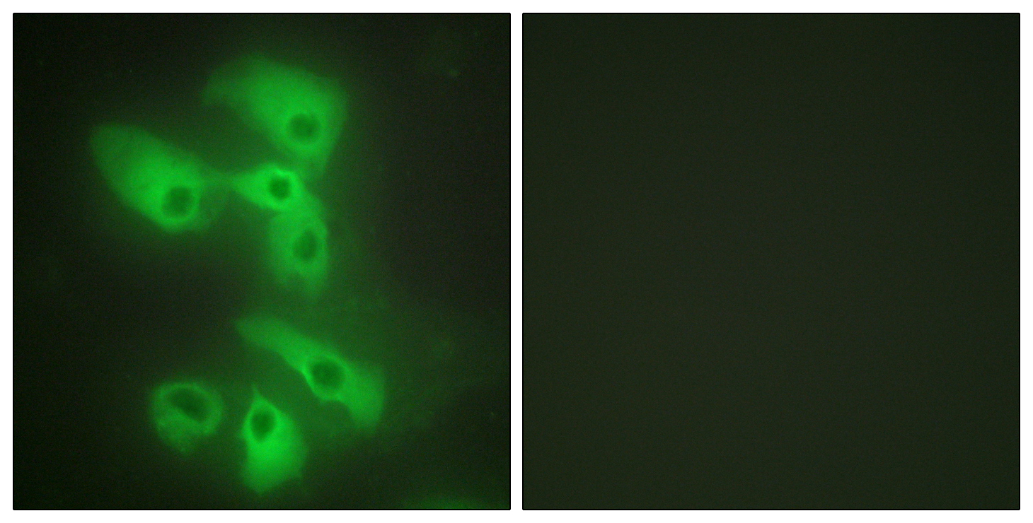
- Immunofluorescence analysis of HeLa cells, using JIP2 Antibody. The picture on the right is blocked with the synthesized peptide.

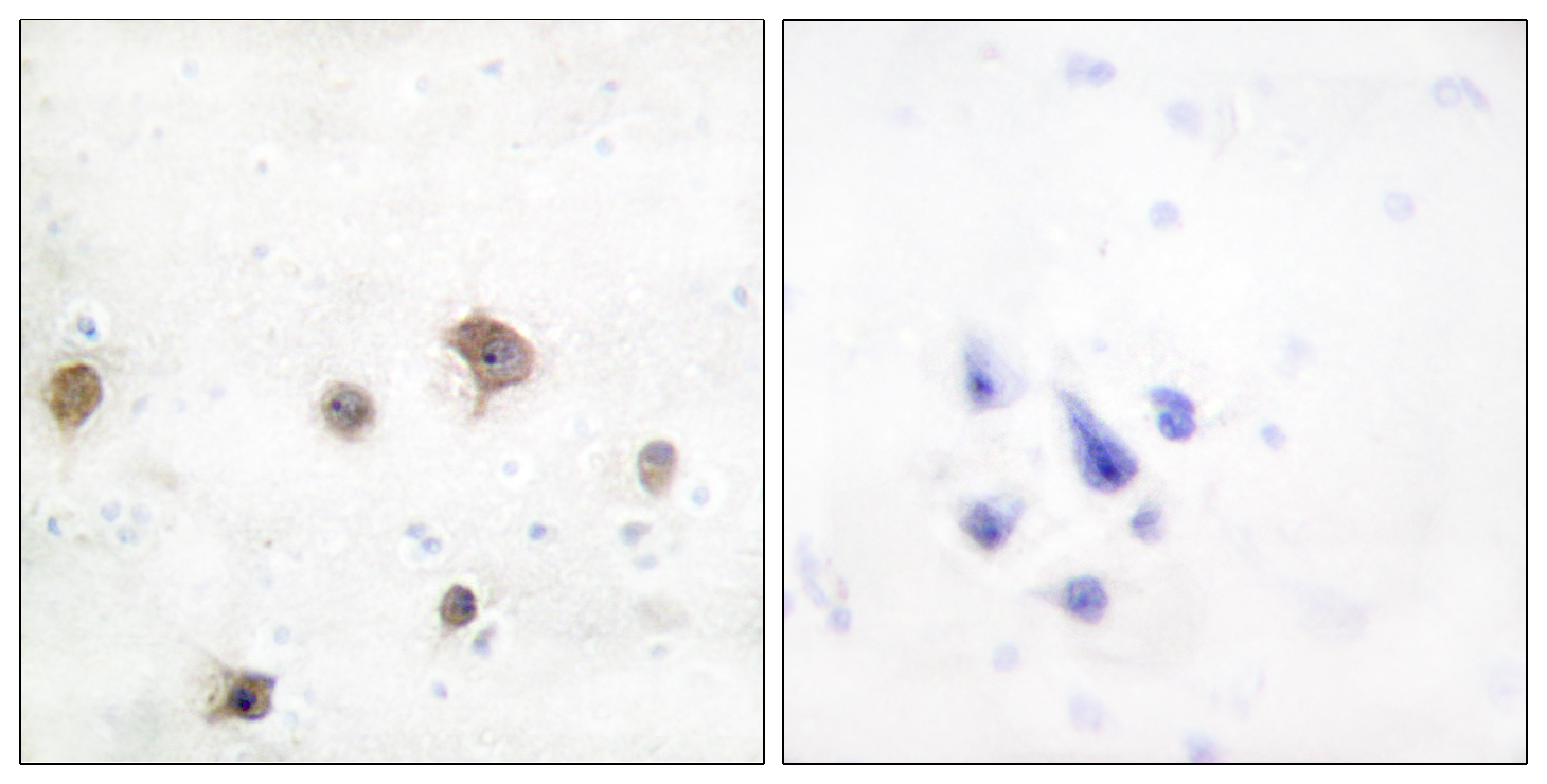
- Immunohistochemistry analysis of paraffin-embedded human brain tissue, using JIP2 Antibody. The picture on the right is blocked with the synthesized peptide.

- Western blot analysis of lysates from mouse brain, using JIP2 Antibody. The lane on the right is blocked with the synthesized peptide.


