IK Polyclonal Antibody
- Catalog No.:YT2297
- Applications:WB;IHC;IF;ELISA
- Reactivity:Human;Mouse;Rat
- Target:
- IK
- Gene Name:
- IK
- Protein Name:
- Protein Red
- Human Gene Id:
- 3550
- Human Swiss Prot No:
- Q13123
- Mouse Gene Id:
- 24010
- Mouse Swiss Prot No:
- Q9Z1M8
- Rat Gene Id:
- 291659
- Rat Swiss Prot No:
- Q66HG8
- Immunogen:
- The antiserum was produced against synthesized peptide derived from human RED. AA range:508-557
- Specificity:
- IK Polyclonal Antibody detects endogenous levels of IK protein.
- Formulation:
- Liquid in PBS containing 50% glycerol, 0.5% BSA and 0.02% sodium azide.
- Source:
- Polyclonal, Rabbit,IgG
- Dilution:
- WB 1:500 - 1:2000. IHC 1:100 - 1:300. ELISA: 1:20000.. IF 1:50-200
- Purification:
- The antibody was affinity-purified from rabbit antiserum by affinity-chromatography using epitope-specific immunogen.
- Concentration:
- 1 mg/ml
- Storage Stability:
- -15°C to -25°C/1 year(Do not lower than -25°C)
- Other Name:
- IK;RED;RER;Protein Red;Cytokine IK;IK factor;Protein RER
- Observed Band(KD):
- 66kD
- Background:
- The protein encoded by this gene was identified by its RED repeat, a stretch of repeated arginine, glutamic acid and aspartic acid residues. The protein localizes to discrete dots within the nucleus, excluding the nucleolus. Its function is unknown. This gene maps to chromosome 5; however, a pseudogene may exist on chromosome 2. [provided by RefSeq, Jul 2008],
- Function:
- caution:Was originally (PubMed:7970704) thought to be the IK factor, a cytokine involved in the negative regulatory pathway of constitutive MHC class II antigens expression.,developmental stage:Expressed at similar levels in fetal and adult tissues.,function:Not known. May bind to chromatin.,PTM:Phosphorylated upon DNA damage, probably by ATM or ATR.,similarity:Belongs to the RED family.,tissue specificity:Ubiquitous.,
- Subcellular Location:
- Nucleus . Nucleus, nucleoplasm . Chromosome . Cytoplasm, cytoskeleton, spindle pole . Predominantly present throughout the nucleoplasm during prometaphase, metaphase and anaphase. Is also detected in nuclear foci that are not identical with Cajal bodies. Starts to accumulate at chromosomes during telophase, and is nearly exclusively associated with chromosomes in newly divided cells (PubMed:24252166). Colocalizes with MAD1L1 at mitotic spindle poles during metaphase and anaphase (PubMed:22351768). .
- Expression:
- Ubiquitous.
- June 19-2018
- WESTERN IMMUNOBLOTTING PROTOCOL
- June 19-2018
- IMMUNOHISTOCHEMISTRY-PARAFFIN PROTOCOL
- June 19-2018
- IMMUNOFLUORESCENCE PROTOCOL
- September 08-2020
- FLOW-CYTOMEYRT-PROTOCOL
- May 20-2022
- Cell-Based ELISA│解您多样本WB检测之困扰
- July 13-2018
- CELL-BASED-ELISA-PROTOCOL-FOR-ACETYL-PROTEIN
- July 13-2018
- CELL-BASED-ELISA-PROTOCOL-FOR-PHOSPHO-PROTEIN
- July 13-2018
- Antibody-FAQs
- Products Images

- Western Blot analysis of various cells using IK Polyclonal Antibody diluted at 1:2000 cells nucleus extracted by Minute TM Cytoplasmic and Nuclear Fractionation kit (SC-003,Inventbiotech,MN,USA).
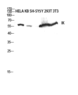
- Western blot analysis of HELA KB SH-SY5Y 293T 3T3 lysis using IK antibody. Antibody was diluted at 1:2000 cells nucleus extracted by Minute TM Cytoplasmic and Nuclear Fractionation kit (SC-003,Inventbiotech,MN,USA).
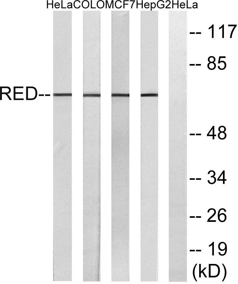
- Western blot analysis of lysates from HepG2, MCF-7, COLO, and HeLa cells, using RED Antibody. The lane on the right is blocked with the synthesized peptide.
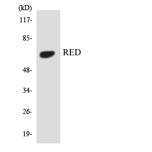
- Western blot analysis of the lysates from 293 cells using RED antibody.
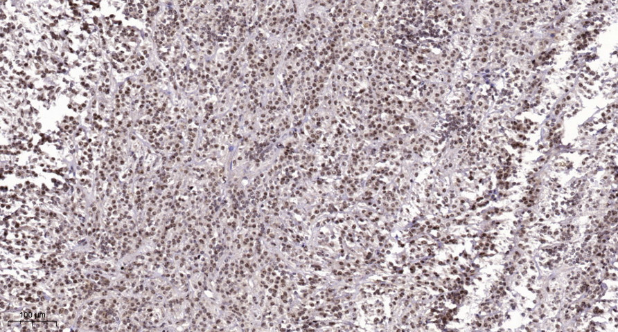
- Immunohistochemical analysis of paraffin-embedded human Colon cancer. 1, Antibody was diluted at 1:200(4° overnight). 2, Tris-EDTA,pH9.0 was used for antigen retrieval. 3,Secondary antibody was diluted at 1:200(room temperature, 45min).


