Histone H2A.X Polyclonal Antibody
- Catalog No.:YT2155
- Applications:WB;IHC;IF;CoIP;ELISA
- Reactivity:Human;Mouse;Rat
- Target:
- Histone H2A.X
- Fields:
- >>Necroptosis;>>Neutrophil extracellular trap formation;>>Alcoholism;>>Systemic lupus erythematosus
- Gene Name:
- H2AFX
- Protein Name:
- Histone H2A.x
- Human Gene Id:
- 3014
- Human Swiss Prot No:
- P16104
- Mouse Gene Id:
- 15270
- Mouse Swiss Prot No:
- P27661
- Immunogen:
- The antiserum was produced against synthesized peptide derived from human Histone H2A.X. AA range:94-143
- Specificity:
- Histone H2A.X Polyclonal Antibody detects endogenous levels of Histone H2A.X protein.
- Formulation:
- Liquid in PBS containing 50% glycerol, 0.5% BSA and 0.02% sodium azide.
- Source:
- Polyclonal, Rabbit,IgG
- Dilution:
- WB 1:500 - 1:2000. IHC 1:100 - 1:300. IF 1:200 - 1:1000. IP 1:200-500,ELISA: 1:10000. Not yet tested in other applications.
- Purification:
- The antibody was affinity-purified from rabbit antiserum by affinity-chromatography using epitope-specific immunogen.
- Concentration:
- 1 mg/ml
- Storage Stability:
- -15°C to -25°C/1 year(Do not lower than -25°C)
- Other Name:
- H2AFX;H2AX;Histone H2A.x;H2a/x
- Observed Band(KD):
- 19kD
- Background:
- Histones are basic nuclear proteins that are responsible for the nucleosome structure of the chromosomal fiber in eukaryotes. Two molecules of each of the four core histones (H2A, H2B, H3, and H4) form an octamer, around which approximately 146 bp of DNA is wrapped in repeating units, called nucleosomes. The linker histone, H1, interacts with linker DNA between nucleosomes and functions in the compaction of chromatin into higher order structures. This gene encodes a replication-independent histone that is a member of the histone H2A family, and generates two transcripts through the use of the conserved stem-loop termination motif, and the polyA addition motif. [provided by RefSeq, Oct 2015],
- Function:
- developmental stage:Synthesized in G1 as well as in S-phase.,domain:The [ST]-Q motif constitutes a recognition sequence for kinases from the PI3/PI4-kinase family.,function:Variant histone H2A which replaces conventional H2A in a subset of nucleosomes. Nucleosomes wrap and compact DNA into chromatin, limiting DNA accessibility to the cellular machineries which require DNA as a template. Histones thereby play a central role in transcription regulation, DNA repair, DNA replication and chromosomal stability. DNA accessibility is regulated via a complex set of post-translational modifications of histones, also called histone code, and nucleosome remodeling. Required for checkpoint-mediated arrest of cell cycle progression in response to low doses of ionizing radiation and for efficient repair of DNA double strand breaks (DSBs) specifically when modified by C-terminal phosphorylation.,PTM:Mon
- Subcellular Location:
- Nucleus . Chromosome .
- Expression:
- Lung,Placenta,
Near-infrared upconversion multimodal nanoparticles for targeted radionuclide therapy of breast cancer lymphatic metastases. Frontiers in Immunology Chuan Zhang, Yujuan Zhang, Maolin Liang, Xiumin Shi, Yan Jun, Longfei Fan, Kai Yang, Feng Wang, Wei Li, Ran Zhu WB Human SKBR3 cell
A PARylation-phosphorylation cascade promotes TOPBP1 loading and RPA-RAD51 exchange in homologous recombination CoIP Human /HeLa cell,U2OS cell
Disrupting PHF8-TOPBP1 connection elicits a breast tumor-specific vulnerability to chemotherapeutics Cancer Lett. 2022 Apr;530:29. WB,IP Human
Marine bromophenol bis (2, 3-dibromo-4, 5-dihydroxybenzyl) ether, represses angiogenesis in HUVEC cells and in zebrafish embryos via inhibiting the VEGF signal systems." Biomedicine & Pharmacotherapy 75 (2015): 58-66.
- June 19-2018
- WESTERN IMMUNOBLOTTING PROTOCOL
- June 19-2018
- IMMUNOHISTOCHEMISTRY-PARAFFIN PROTOCOL
- June 19-2018
- IMMUNOFLUORESCENCE PROTOCOL
- September 08-2020
- FLOW-CYTOMEYRT-PROTOCOL
- May 20-2022
- Cell-Based ELISA│解您多样本WB检测之困扰
- July 13-2018
- CELL-BASED-ELISA-PROTOCOL-FOR-ACETYL-PROTEIN
- July 13-2018
- CELL-BASED-ELISA-PROTOCOL-FOR-PHOSPHO-PROTEIN
- July 13-2018
- Antibody-FAQs
- Products Images
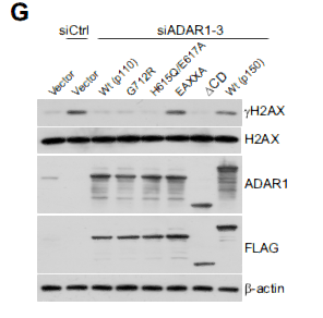
- ADAR1 links R-loop homeostasis to ATR activation in replication stress response. NUCLEIC ACIDS RESEARCH Lei Shi WB,CoIP Human HeLa cell
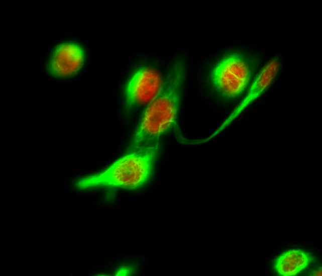
- Immunofluorescence analysis of Hela cell. 1,Histone H2A.X Polyclonal Antibody(red) was diluted at 1:200(4° overnight). LC3B Polyclonal Antibody(green) was diluted at 1:200(4° overnight). 2, Goat Anti Rabbit Alexa Fluor 594 Catalog:RS3611 was diluted at 1:1000(room temperature, 50min). Goat Anti Mouse Alexa Fluor 488 Catalog:RS3208 was diluted at 1:1000(room temperature, 50min).
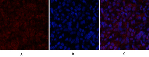
- Immunofluorescence analysis of rat-lung tissue. 1,Histone H2A.X Polyclonal Antibody(red) was diluted at 1:200(4°C,overnight). 2, Cy3 labled Secondary antibody was diluted at 1:300(room temperature, 50min).3, Picture B: DAPI(blue) 10min. Picture A:Target. Picture B: DAPI. Picture C: merge of A+B
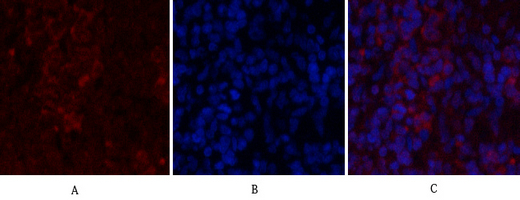
- Immunofluorescence analysis of rat-spleen tissue. 1,Histone H2A.X Polyclonal Antibody(red) was diluted at 1:200(4°C,overnight). 2, Cy3 labled Secondary antibody was diluted at 1:300(room temperature, 50min).3, Picture B: DAPI(blue) 10min. Picture A:Target. Picture B: DAPI. Picture C: merge of A+B

- Immunohistochemical analysis of paraffin-embedded Human-uterus tissue. 1,Histone H2A.X Polyclonal Antibody was diluted at 1:200(4°C,overnight). 2, Sodium citrate pH 6.0 was used for antibody retrieval(>98°C,20min). 3,Secondary antibody was diluted at 1:200(room tempeRature, 30min). Negative control was used by secondary antibody only.
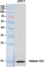
- Western Blot analysis of various cells using Histone H2A.X Polyclonal Antibody diluted at 1:2000 cells nucleus extracted by Minute TM Cytoplasmic and Nuclear Fractionation kit (SC-003,Inventbiotech,MN,USA).
.jpg)
- Western Blot analysis of HEPG2 cells using Histone H2A.X Polyclonal Antibody diluted at 1:2000 cells nucleus extracted by Minute TM Cytoplasmic and Nuclear Fractionation kit (SC-003,Inventbiotech,MN,USA).
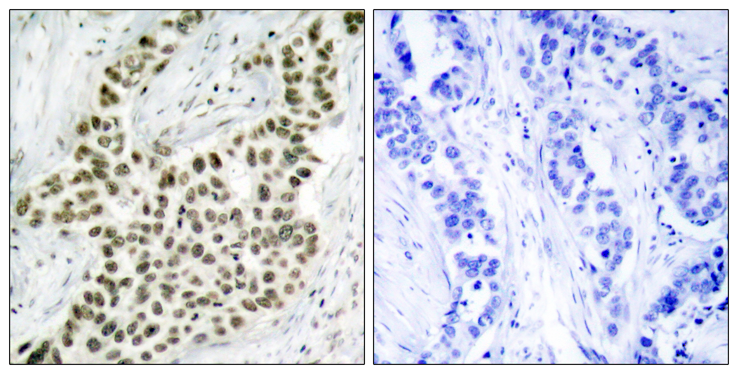
- Immunohistochemistry analysis of paraffin-embedded human breast carcinoma tissue, using Histone H2A.X Antibody. The picture on the right is blocked with the synthesized peptide.


