HDAC6 Polyclonal Antibody
- Catalog No.:YT2118
- Applications:WB;IHC;IF;ELISA
- Reactivity:Human;Mouse
- Target:
- HDAC6
- Fields:
- >>Neutrophil extracellular trap formation;>>Amyotrophic lateral sclerosis;>>Alcoholism;>>Viral carcinogenesis
- Gene Name:
- HDAC6
- Protein Name:
- Histone deacetylase 6
- Human Gene Id:
- 10013
- Human Swiss Prot No:
- Q9UBN7
- Mouse Gene Id:
- 15185
- Mouse Swiss Prot No:
- Q9Z2V5
- Immunogen:
- The antiserum was produced against synthesized peptide derived from human HDAC6. AA range:7-56
- Specificity:
- HDAC6 Polyclonal Antibody detects endogenous levels of HDAC6 protein.
- Formulation:
- Liquid in PBS containing 50% glycerol, 0.5% BSA and 0.02% sodium azide.
- Source:
- Polyclonal, Rabbit,IgG
- Dilution:
- WB 1:500 - 1:2000. IHC 1:100 - 1:300. IF 1:200 - 1:1000. ELISA: 1:20000. Not yet tested in other applications.
- Purification:
- The antibody was affinity-purified from rabbit antiserum by affinity-chromatography using epitope-specific immunogen.
- Concentration:
- 1 mg/ml
- Storage Stability:
- -15°C to -25°C/1 year(Do not lower than -25°C)
- Other Name:
- HDAC6;KIAA0901;JM21;Histone deacetylase 6;HD6
- Observed Band(KD):
- 131kD
- Background:
- Histones play a critical role in transcriptional regulation, cell cycle progression, and developmental events. Histone acetylation/deacetylation alters chromosome structure and affects transcription factor access to DNA. The protein encoded by this gene belongs to class II of the histone deacetylase/acuc/apha family. It contains an internal duplication of two catalytic domains which appear to function independently of each other. This protein possesses histone deacetylase activity and represses transcription. [provided by RefSeq, Jul 2008],
- Function:
- catalytic activity:Hydrolysis of an N(6)-acetyl-lysine residue of a histone to yield a deacetylated histone.,function:Responsible for the deacetylation of lysine residues on the N-terminal part of the core histones (H2A, H2B, H3 and H4). Histone deacetylation gives a tag for epigenetic repression and plays an important role in transcriptional regulation, cell cycle progression and developmental events. Histone deacetylases act via the formation of large multiprotein complexes (By similarity). Plays a central role in microtubule-dependent cell motility via deacetylation of tubulin.,PTM:Sumoylated in vitro.,PTM:Ubiquitinated. Its polyubiquitination however does not lead to its degradation.,similarity:Belongs to the histone deacetylase family. Type 2 subfamily.,similarity:Contains 1 UBP-type zinc finger.,subcellular location:It is mainly cytoplasmic, where it is associated with microtubules
- Subcellular Location:
- Cytoplasm . Cytoplasm, cytoskeleton . Nucleus . Perikaryon . Cell projection, dendrite . Cell projection, axon . It is mainly cytoplasmic, where it is associated with microtubules. .
- Expression:
- Brain,Epithelium,Kidney,Muscle,Ovary,Placenta,
Deacetylation of tumor-suppressor MST1 in Hippo pathway induces its degradation through HBXIP-elevated HDAC6 in promotion of breast cancer growth. ONCOGENE 2015 Dec 14 WB Human MCF-7 cell,HEK293T cell
HDAC-selective Inhibitor Cay10603 Has Single Anti-tumour Effect in Burkitt’s Lymphoma Cells by Impeding the Cell Cycle. Current Medical Science 2019 Aug 05 WB Human CA46 cell, Raji Cell
- June 19-2018
- WESTERN IMMUNOBLOTTING PROTOCOL
- June 19-2018
- IMMUNOHISTOCHEMISTRY-PARAFFIN PROTOCOL
- June 19-2018
- IMMUNOFLUORESCENCE PROTOCOL
- September 08-2020
- FLOW-CYTOMEYRT-PROTOCOL
- May 20-2022
- Cell-Based ELISA│解您多样本WB检测之困扰
- July 13-2018
- CELL-BASED-ELISA-PROTOCOL-FOR-ACETYL-PROTEIN
- July 13-2018
- CELL-BASED-ELISA-PROTOCOL-FOR-PHOSPHO-PROTEIN
- July 13-2018
- Antibody-FAQs
- Products Images
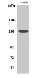
- Western Blot analysis of various cells using HDAC6 Polyclonal Antibody diluted at 1:1000
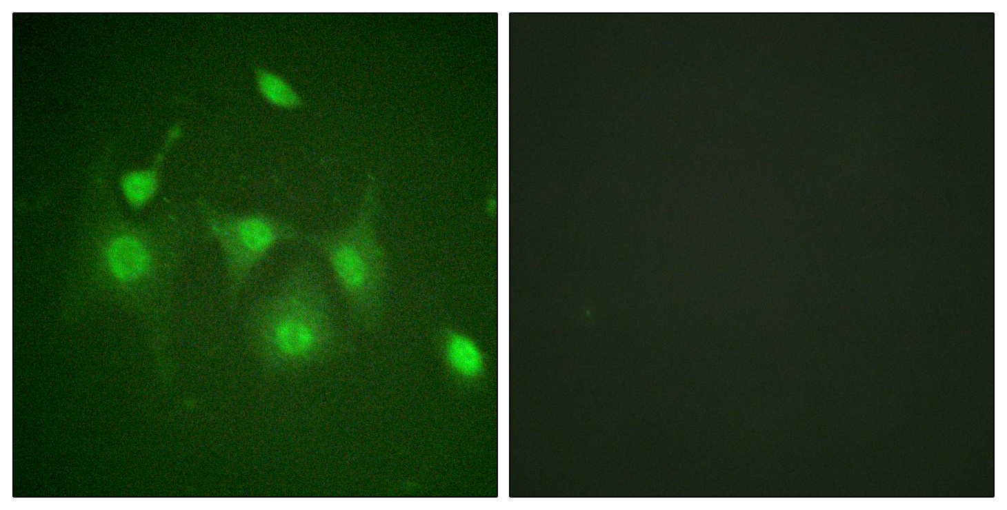
- Immunofluorescence analysis of HepG2 cells, using HDAC6 Antibody. The picture on the right is blocked with the synthesized peptide.
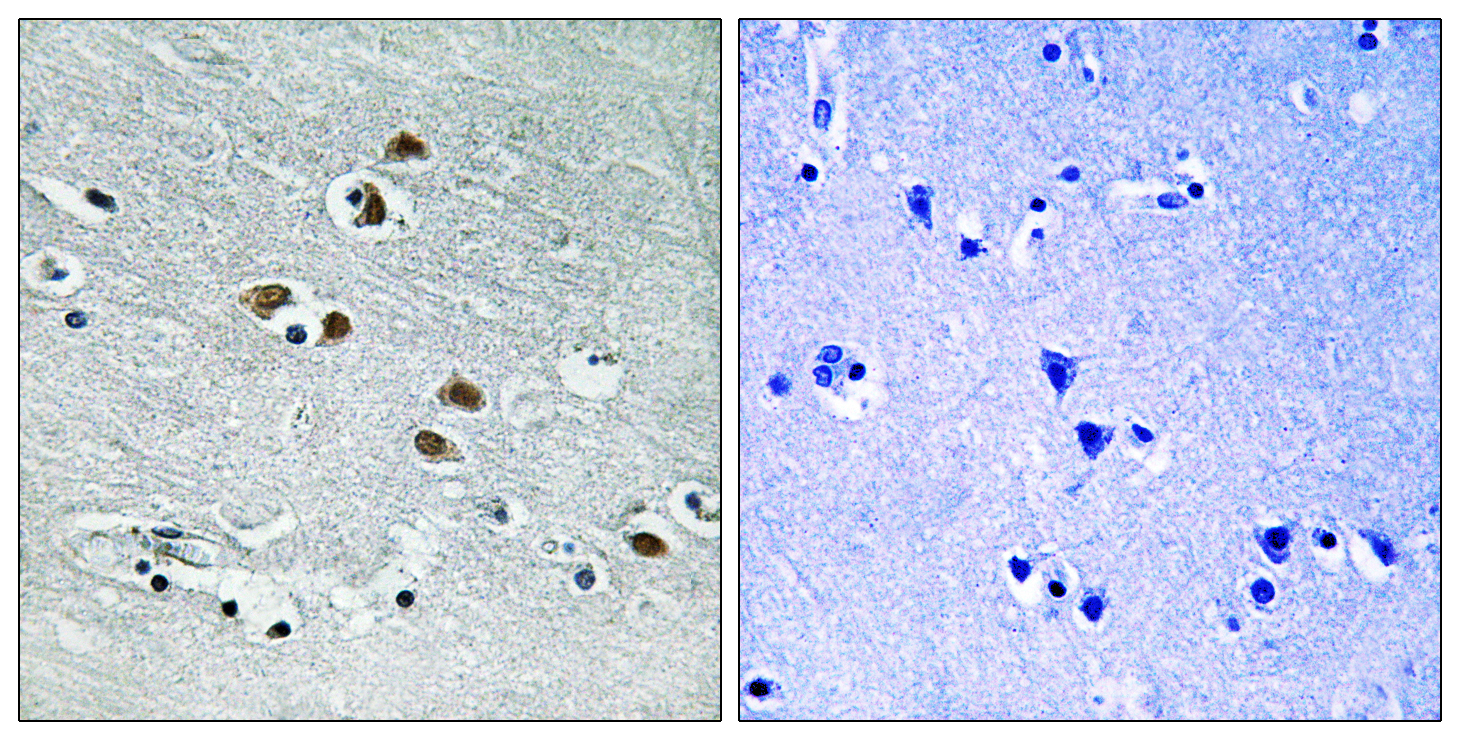
- Immunohistochemistry analysis of paraffin-embedded human brain tissue, using HDAC6 Antibody. The picture on the right is blocked with the synthesized peptide.
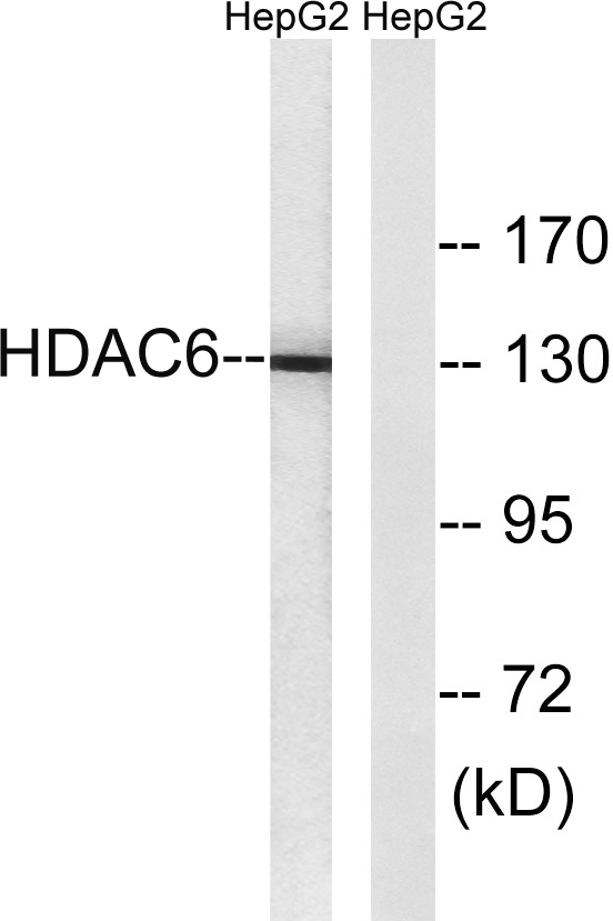
- Western blot analysis of lysates from HepG2 cells, using HDAC6 Antibody. The lane on the right is blocked with the synthesized peptide.


