HCCS Polyclonal Antibody
- Catalog No.:YT2107
- Applications:WB;IHC;IF;ELISA
- Reactivity:Human;Mouse;Monkey
- Target:
- HCCS
- Fields:
- >>Porphyrin metabolism;>>Metabolic pathways
- Gene Name:
- HCCS
- Protein Name:
- Cytochrome c-type heme lyase
- Human Gene Id:
- 3052
- Human Swiss Prot No:
- P53701
- Mouse Gene Id:
- 15159
- Mouse Swiss Prot No:
- P53702
- Immunogen:
- The antiserum was produced against synthesized peptide derived from human Cytochrome c-type Heme Lyase. AA range:81-130
- Specificity:
- HCCS Polyclonal Antibody detects endogenous levels of HCCS protein.
- Formulation:
- Liquid in PBS containing 50% glycerol, 0.5% BSA and 0.02% sodium azide.
- Source:
- Polyclonal, Rabbit,IgG
- Dilution:
- WB 1:500 - 1:2000. IHC 1:100 - 1:300. IF 1:200 - 1:1000. ELISA: 1:20000. Not yet tested in other applications.
- Purification:
- The antibody was affinity-purified from rabbit antiserum by affinity-chromatography using epitope-specific immunogen.
- Concentration:
- 1 mg/ml
- Storage Stability:
- -15°C to -25°C/1 year(Do not lower than -25°C)
- Other Name:
- HCCS;CCHL;Cytochrome c-type heme lyase;CCHL;Holocytochrome c-type synthase
- Observed Band(KD):
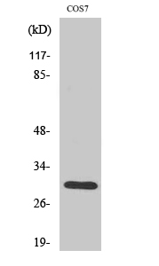
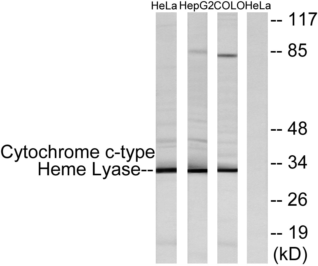
- 31kD
- Background:
- holocytochrome c synthase(HCCS) Homo sapiens The protein encoded by this gene is an enzyme that covalently links a heme group to the apoprotein of cytochrome c. Defects in this gene are a cause of microphthalmia syndromic type 7 (MCOPS7). Three transcript variants encoding the same protein have been found for this gene. [provided by RefSeq, Jan 2010],
- Function:
- catalytic activity:Holocytochrome c = apocytochrome c + heme.,disease:Defects in HCCS are a cause of microphthalmia syndromic type 7 (MCOPS7) [MIM:309801]; also known as microphthalmia with linear skin defects (MLS) or MIDAS syndrome. Microphthalmia is a clinically heterogeneous disorder of eye formation, ranging from small size of a single eye TO complete bilateral absence of ocular tissues (anophthalmia). In many cases, microphthalmia/anophthalmia occurs in association with syndromes that include non-ocular abnormalities. MCOPS7 is a disorder characterized by unilateral or bilateral microphthalmia, linear skin defects in affected females, and in utero lethality for males. Skin defects are limited to the face and neck, consisting of areas of aplastic skin that heal with age to form hyperpigmented areas. Additional features in female patients include agenesis of the corpus callosum, scle
- Subcellular Location:
- Mitochondrion inner membrane . Membrane ; Lipid-anchor .
- Expression:
- Brain,Liver,Ovary,
- June 19-2018
- WESTERN IMMUNOBLOTTING PROTOCOL
- June 19-2018
- IMMUNOHISTOCHEMISTRY-PARAFFIN PROTOCOL
- June 19-2018
- IMMUNOFLUORESCENCE PROTOCOL
- September 08-2020
- FLOW-CYTOMEYRT-PROTOCOL
- May 20-2022
- Cell-Based ELISA│解您多样本WB检测之困扰
- July 13-2018
- CELL-BASED-ELISA-PROTOCOL-FOR-ACETYL-PROTEIN
- July 13-2018
- CELL-BASED-ELISA-PROTOCOL-FOR-PHOSPHO-PROTEIN
- July 13-2018
- Antibody-FAQs
- Products Images

- Western Blot analysis of various cells using HCCS Polyclonal Antibody diluted at 1:2000

- Immunofluorescence analysis of MCF7 cells, using Cytochrome c-type Heme Lyase Antibody. The picture on the right is blocked with the synthesized peptide.

- Immunohistochemistry analysis of paraffin-embedded human tonsil tissue, using Cytochrome c-type Heme Lyase Antibody. The picture on the right is blocked with the synthesized peptide.

- Western blot analysis of lysates from HeLa, HepG2, and COLO cells, using Cytochrome c-type Heme Lyase Antibody. The lane on the right is blocked with the synthesized peptide.


