GPRC5B Polyclonal Antibody
- Catalog No.:YT2037
- Applications:WB;IF;ELISA
- Reactivity:Human;Rat;Mouse;
- Target:
- GPRC5B
- Gene Name:
- GPRC5B
- Protein Name:
- G-protein coupled receptor family C group 5 member B
- Human Gene Id:
- 51704
- Human Swiss Prot No:
- Q9NZH0
- Mouse Swiss Prot No:
- Q923Z0
- Immunogen:
- The antiserum was produced against synthesized peptide derived from human GPRC5B. AA range:61-110
- Specificity:
- GPRC5B Polyclonal Antibody detects endogenous levels of GPRC5B protein.
- Formulation:
- Liquid in PBS containing 50% glycerol, 0.5% BSA and 0.02% sodium azide.
- Source:
- Polyclonal, Rabbit,IgG
- Dilution:
- WB 1:500 - 1:2000. IF 1:200 - 1:1000. ELISA: 1:5000. Not yet tested in other applications.
- Purification:
- The antibody was affinity-purified from rabbit antiserum by affinity-chromatography using epitope-specific immunogen.
- Concentration:
- 1 mg/ml
- Storage Stability:
- -15°C to -25°C/1 year(Do not lower than -25°C)
- Other Name:
- GPRC5B;RAIG2;G-protein coupled receptor family C group 5 member B;A-69G12.1;Retinoic acid-induced gene 2 protein;RAIG-2
- Observed Band(KD):
- 48kD
- Background:
- This gene encodes a member of the type 3 G protein-coupled receptor family. Members of this superfamily are characterized by a signature 7-transmembrane domain motif. The encoded protein may modulate insulin secretion and increased protein expression is associated with type 2 diabetes. Alternative splicing results in multiple transcript variants. [provided by RefSeq, Feb 2015],
- Function:
- caution:It is uncertain whether Met-1 or Met-9 is the initiator.,function:Unknown. This retinoic acid-inducible G-protein coupled receptor provide evidence for a possible interaction between retinoid and G-protein signaling pathways.,induction:By all-trans retinoic acid (ATRA).,similarity:Belongs to the G-protein coupled receptor 3 family.,subcellular location:Localized in the plasma membrane and perinuclear vesicles.,tissue specificity:Expression is high in kidney, pancreas, and testis, medium in brain, heart, prostate, small intestine, and spleen, low in liver, placenta, skeletal muscle, colon, ovary, and thymus, and not detectable in lung and peripheral leukocyte. According to PubMed:10945465: highly expressed in most brain areas examined, with the highest levels observed in corpus callosum, caudate nucleus, putamen, substantia nigra, thalamus, hippocampus, and spinal chord as well as
- Subcellular Location:
- Cell membrane ; Multi-pass membrane protein . Cytoplasmic vesicle membrane ; Multi-pass membrane protein . Localized in the plasma membrane and perinuclear vesicles.
- Expression:
- Expression is high in kidney, pancreas, and testis, medium in brain, heart, prostate, small intestine, and spleen, low in liver, placenta, skeletal muscle, colon, ovary, and thymus, and not detectable in lung and peripheral leukocyte. According to PubMed:10945465, highly expressed in most brain areas examined, with the highest levels observed in corpus callosum, caudate nucleus, putamen, substantia nigra, thalamus, hippocampus, and spinal cord as well as in dorsal root ganglia (DRG). In the periphery, expression levels are relatively low, compared to the CNS, with the strongest expression detected in pancreas, testis, uterus, and stomach.
- June 19-2018
- WESTERN IMMUNOBLOTTING PROTOCOL
- June 19-2018
- IMMUNOHISTOCHEMISTRY-PARAFFIN PROTOCOL
- June 19-2018
- IMMUNOFLUORESCENCE PROTOCOL
- September 08-2020
- FLOW-CYTOMEYRT-PROTOCOL
- May 20-2022
- Cell-Based ELISA│解您多样本WB检测之困扰
- July 13-2018
- CELL-BASED-ELISA-PROTOCOL-FOR-ACETYL-PROTEIN
- July 13-2018
- CELL-BASED-ELISA-PROTOCOL-FOR-PHOSPHO-PROTEIN
- July 13-2018
- Antibody-FAQs
- Products Images
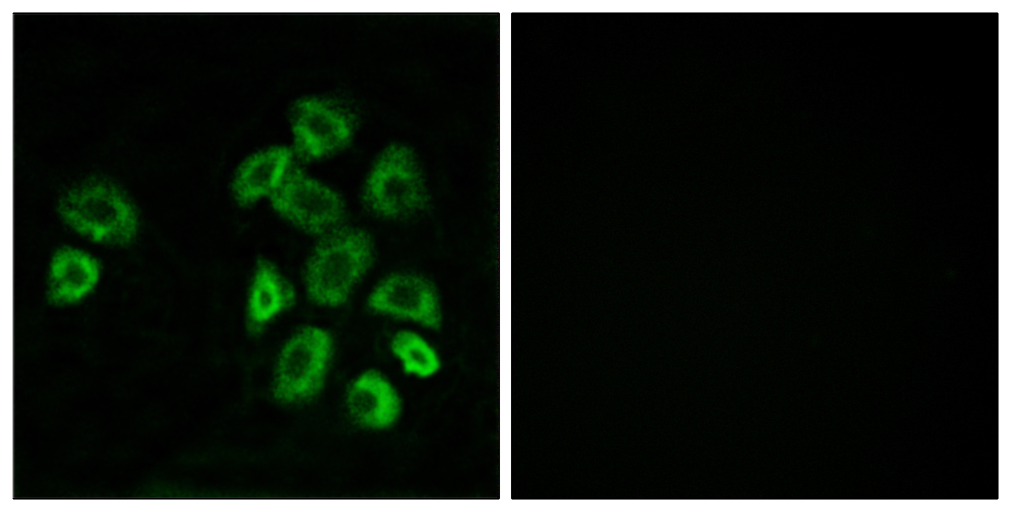
- Immunofluorescence analysis of MCF7 cells, using GPRC5B Antibody. The picture on the right is blocked with the synthesized peptide.
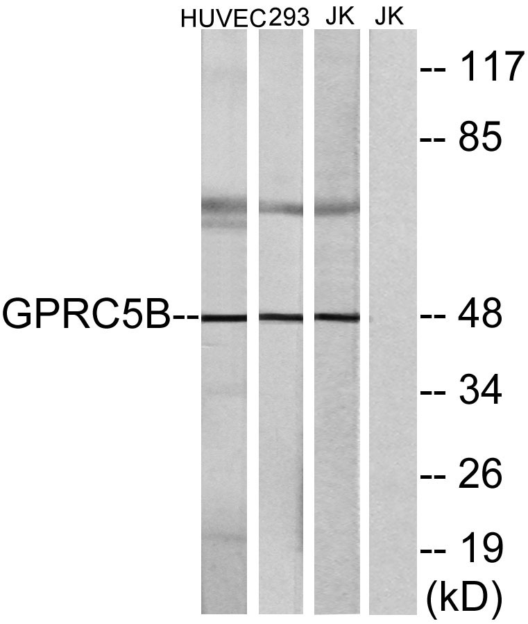
- Western blot analysis of lysates from Jurkat, HUVEC, and 293 cells, using GPRC5B Antibody. The lane on the right is blocked with the synthesized peptide.


