GPR56 Polyclonal Antibody
- Catalog No.:YT2026
- Applications:WB;IF;ELISA
- Reactivity:Human;Rat;Mouse;
- Target:
- GPR56
- Gene Name:
- GPR56
- Protein Name:
- G-protein coupled receptor 56
- Human Gene Id:
- 9289
- Human Swiss Prot No:
- Q9Y653
- Mouse Swiss Prot No:
- Q8K209
- Immunogen:
- The antiserum was produced against synthesized peptide derived from human GPR56. AA range:251-300
- Specificity:
- GPR56 Polyclonal Antibody detects endogenous levels of GPR56 protein.
- Formulation:
- Liquid in PBS containing 50% glycerol, 0.5% BSA and 0.02% sodium azide.
- Source:
- Polyclonal, Rabbit,IgG
- Dilution:
- WB 1:500 - 1:2000. IF 1:200 - 1:1000. ELISA: 1:20000. Not yet tested in other applications.
- Purification:
- The antibody was affinity-purified from rabbit antiserum by affinity-chromatography using epitope-specific immunogen.
- Concentration:
- 1 mg/ml
- Storage Stability:
- -15°C to -25°C/1 year(Do not lower than -25°C)
- Other Name:
- GPR56;TM7LN4;TM7XN1;G-protein coupled receptor 56;Protein TM7XN1
- Observed Band(KD):
- 78kD
- Background:
- This gene encodes a member of the G protein-coupled receptor family and regulates brain cortical patterning. The encoded protein binds specifically to transglutaminase 2, a component of tissue and tumor stroma implicated as an inhibitor of tumor progression. Mutations in this gene are associated with a brain malformation known as bilateral frontoparietal polymicrogyria. Alternative splicing results in multiple transcript variants. [provided by RefSeq, Feb 2014],
- Function:
- disease:Defects in GPR56 are the cause of bilateral frontoparietal polymicrogyria (BFPP) [MIM:606854]. BFPP is characterized by disorganized cortical lamination that is most severe in frontal cortex.,function:Could be involved in cell-cell interactions.,similarity:Belongs to the G-protein coupled receptor 2 family. LN-TM7 subfamily.,similarity:Contains 1 GPS domain.,tissue specificity:Widely distributed with highest levels found in thyroid gland, brain and heart. Expressed in a great number of tumor cells.,
- Subcellular Location:
- Cell membrane ; Multi-pass membrane protein .; [ADGRG1 N-terminal fragment]: Secreted .; [ADGRG1 C-terminal fragment]: Membrane raft . Interaction with its ligand COL3A1 leads to the release of ADGRG1 NT from the membrane and triggers the association of ADGRG1 CT with lipid rafts. .
- Expression:
- Widely distributed with highest levels found in thyroid gland, brain and heart. Expressed in a great number of tumor cells. Expression is down-regulated in different tumors from highly metastatic cells.
- June 19-2018
- WESTERN IMMUNOBLOTTING PROTOCOL
- June 19-2018
- IMMUNOHISTOCHEMISTRY-PARAFFIN PROTOCOL
- June 19-2018
- IMMUNOFLUORESCENCE PROTOCOL
- September 08-2020
- FLOW-CYTOMEYRT-PROTOCOL
- May 20-2022
- Cell-Based ELISA│解您多样本WB检测之困扰
- July 13-2018
- CELL-BASED-ELISA-PROTOCOL-FOR-ACETYL-PROTEIN
- July 13-2018
- CELL-BASED-ELISA-PROTOCOL-FOR-PHOSPHO-PROTEIN
- July 13-2018
- Antibody-FAQs
- Products Images
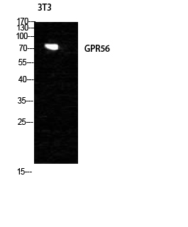
- Western Blot analysis of NIH-3T3 cells using GPR56 Polyclonal Antibody diluted at 1:1000
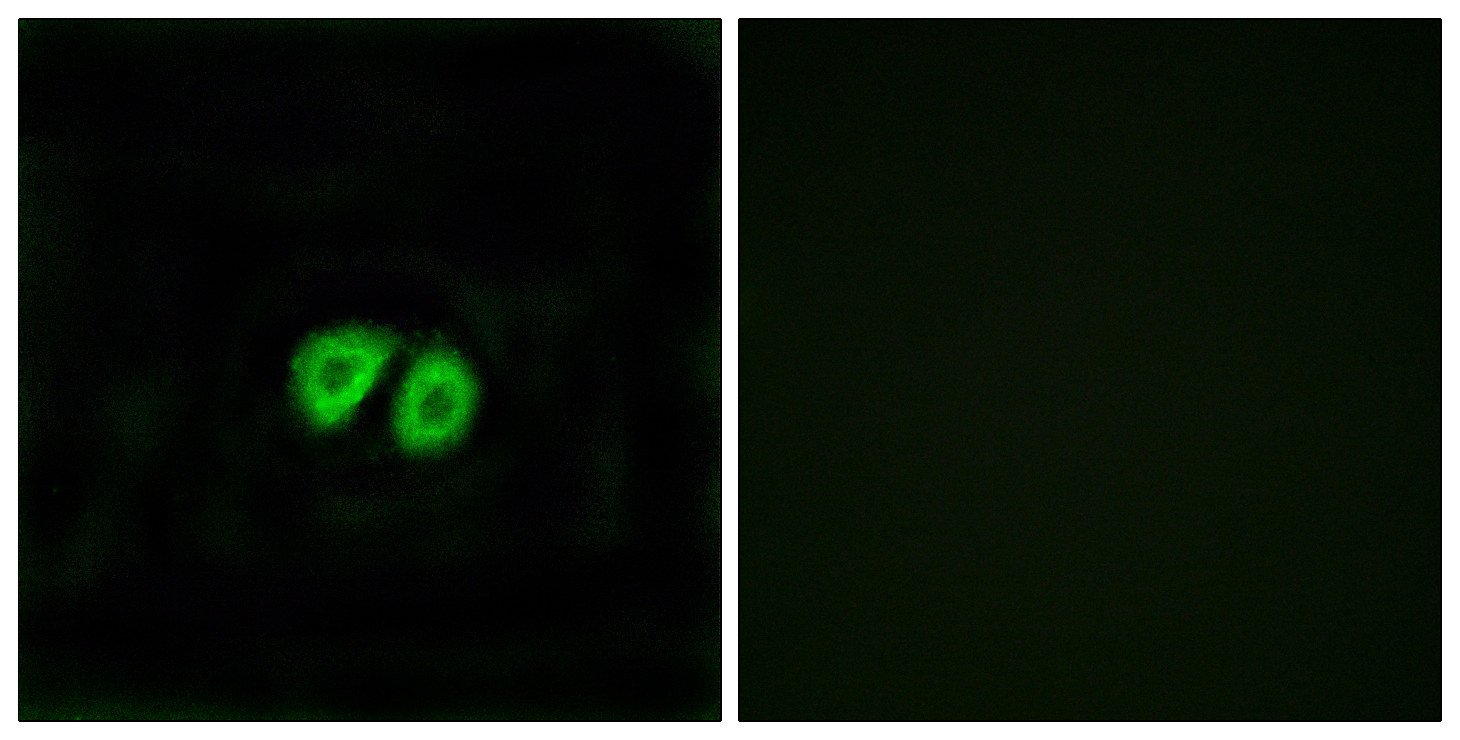
- Immunofluorescence analysis of MCF7 cells, using GPR56 Antibody. The picture on the right is blocked with the synthesized peptide.
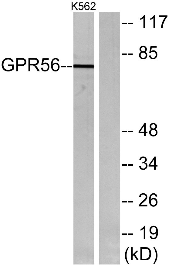
- Western blot analysis of lysates from K562 cells, using GPR56 Antibody. The lane on the right is blocked with the synthesized peptide.


