GPR41 Polyclonal Antibody
- Catalog No.:YT2020
- Applications:WB;IF;ELISA
- Reactivity:Human;Mouse
- Target:
- GPR41
- Gene Name:
- FFAR3
- Protein Name:
- Free fatty acid receptor 3
- Human Gene Id:
- 2865
- Human Swiss Prot No:
- O14843
- Mouse Gene Id:
- 233080
- Mouse Swiss Prot No:
- Q3UFD7
- Immunogen:
- The antiserum was produced against synthesized peptide derived from human FFAR3. AA range:11-60
- Specificity:
- GPR41 Polyclonal Antibody detects endogenous levels of GPR41 protein.
- Formulation:
- Liquid in PBS containing 50% glycerol, 0.5% BSA and 0.02% sodium azide.
- Source:
- Polyclonal, Rabbit,IgG
- Dilution:
- WB 1:500 - 1:2000. IF 1:200 - 1:1000. ELISA: 1:5000. Not yet tested in other applications.
- Purification:
- The antibody was affinity-purified from rabbit antiserum by affinity-chromatography using epitope-specific immunogen.
- Concentration:
- 1 mg/ml
- Storage Stability:
- -15°C to -25°C/1 year(Do not lower than -25°C)
- Other Name:
- FFAR3;GPR41;Free fatty acid receptor 3;G-protein coupled receptor 41
- Observed Band(KD):
- 38kD
- Background:
- function:Receptor for short chain fatty acids through a G(i)-protein-mediated inhibition of adenylyl cyclase and elevation of intracellular calcium. The rank order of potency for agonists of this receptor is propionate = pentanoate = butyrate > acetate > formate.,similarity:Belongs to the G-protein coupled receptor 1 family.,tissue specificity:Highest level in adipose tissue, and lower expression across all tissues tested.,
- Function:
- function:Receptor for short chain fatty acids through a G(i)-protein-mediated inhibition of adenylyl cyclase and elevation of intracellular calcium. The rank order of potency for agonists of this receptor is propionate = pentanoate = butyrate > acetate > formate.,similarity:Belongs to the G-protein coupled receptor 1 family.,tissue specificity:Highest level in adipose tissue, and lower expression across all tissues tested.,
- Subcellular Location:
- Cell membrane ; Multi-pass membrane protein .
- Expression:
- Highest level in adipose tissue, and lower expression across all tissues tested. Expressed in sympathetic ganglia.
Bifidobacterium animalis subsp. lactis A6 Enhances Fatty Acid β-Oxidation of Adipose Tissue to Ameliorate the Development of Obesity in Mice Nutrients. 2022 Jan;14(3):598. WB Mouse epididymal adipose tissues
Gut microbiota modulate CD8+ T cell immunity in gastric cancer through Butyrate/GPR109A/HOPX Gut Microbes Xiang Yu WB Human 1:1000 GES-1 cell,MGC-803 cell,HGC-27 cell,SNU-216 cell
- June 19-2018
- WESTERN IMMUNOBLOTTING PROTOCOL
- June 19-2018
- IMMUNOHISTOCHEMISTRY-PARAFFIN PROTOCOL
- June 19-2018
- IMMUNOFLUORESCENCE PROTOCOL
- September 08-2020
- FLOW-CYTOMEYRT-PROTOCOL
- May 20-2022
- Cell-Based ELISA│解您多样本WB检测之困扰
- July 13-2018
- CELL-BASED-ELISA-PROTOCOL-FOR-ACETYL-PROTEIN
- July 13-2018
- CELL-BASED-ELISA-PROTOCOL-FOR-PHOSPHO-PROTEIN
- July 13-2018
- Antibody-FAQs
- Products Images
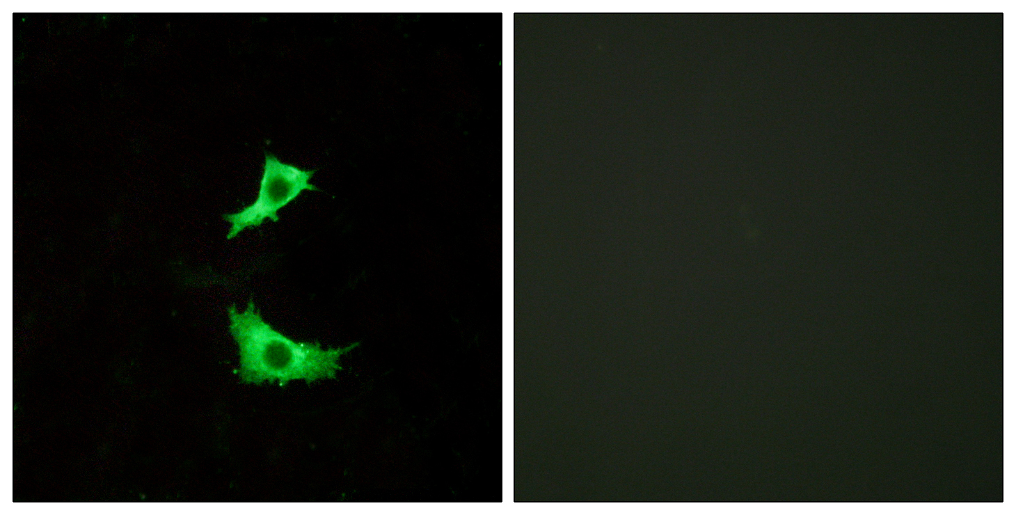
- Immunofluorescence analysis of LOVO cells, using FFAR3 Antibody. The picture on the right is blocked with the synthesized peptide.
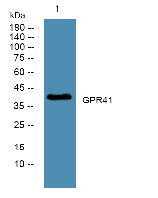
- Western blot analysis of lysates from U2OS cells, primary antibody was diluted at 1:1000, 4°over night
- Bifidobacterium animalis subsp. lactis A6 Enhances Fatty Acid β-Oxidation of Adipose Tissue to Ameliorate the Development of Obesity in Mice Nutrients. 2022 Jan;14(3):598. WB Mouse epididymal adipose tissues


