GPR40 Polyclonal Antibody
- Catalog No.:YT2019
- Applications:WB;IF;ELISA
- Reactivity:Human;Monkey
- Target:
- GPR40
- Fields:
- >>Insulin secretion
- Gene Name:
- FFAR1
- Protein Name:
- Free fatty acid receptor 1
- Human Gene Id:
- 2864
- Human Swiss Prot No:
- O14842
- Mouse Swiss Prot No:
- Q76JU9
- Immunogen:
- The antiserum was produced against synthesized peptide derived from human FFAR1. AA range:185-234
- Specificity:
- GPR40 Polyclonal Antibody detects endogenous levels of GPR40 protein.
- Formulation:
- Liquid in PBS containing 50% glycerol, 0.5% BSA and 0.02% sodium azide.
- Source:
- Polyclonal, Rabbit,IgG
- Dilution:
- WB 1:500 - 1:2000. IF 1:200 - 1:1000. ELISA: 1:10000. Not yet tested in other applications.
- Purification:
- The antibody was affinity-purified from rabbit antiserum by affinity-chromatography using epitope-specific immunogen.
- Concentration:
- 1 mg/ml
- Storage Stability:
- -15°C to -25°C/1 year(Do not lower than -25°C)
- Other Name:
- FFAR1;GPR40;Free fatty acid receptor 1;G-protein coupled receptor 40
- Observed Band(KD):
- 26kD
- Background:
- This gene encodes a member of the GP40 family of G protein-coupled receptors that are clustered together on chromosome 19. The encoded protein is a receptor for medium and long chain free fatty acids and may be involved in the metabolic regulation of insulin secretion. Polymorphisms in this gene may be associated with type 2 diabetes. [provided by RefSeq, Apr 2009],
- Function:
- function:Receptor for medium and long chain saturated and unsaturated fatty acids. Binding of the ligand increase intracellular calcium concentration and amplify glucose-stimulated insulin secretion. The activity of this receptor is mediated by G-proteins that activate phospholipase C. Seems to act through a G(q) and G(i)-mediated pathway.,similarity:Belongs to the G-protein coupled receptor 1 family.,tissue specificity:Expressed abundantly in pancreatic beta cells.,
- Subcellular Location:
- Cell membrane ; Multi-pass membrane protein .
- Expression:
- Detected in brain and pancreas. Detected in pancreatic beta cells.
- June 19-2018
- WESTERN IMMUNOBLOTTING PROTOCOL
- June 19-2018
- IMMUNOHISTOCHEMISTRY-PARAFFIN PROTOCOL
- June 19-2018
- IMMUNOFLUORESCENCE PROTOCOL
- September 08-2020
- FLOW-CYTOMEYRT-PROTOCOL
- May 20-2022
- Cell-Based ELISA│解您多样本WB检测之困扰
- July 13-2018
- CELL-BASED-ELISA-PROTOCOL-FOR-ACETYL-PROTEIN
- July 13-2018
- CELL-BASED-ELISA-PROTOCOL-FOR-PHOSPHO-PROTEIN
- July 13-2018
- Antibody-FAQs
- Products Images
.jpg)
- Western Blot analysis of COS7 cells using GPR40 Polyclonal Antibody diluted at 1:500
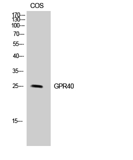
- Western Blot analysis of COS-7 cells using GPR40 Polyclonal Antibody diluted at 1:500
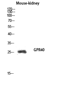
- Western blot analysis of Mouse-kidney lysis using GPR40 antibody. Antibody was diluted at 1:500
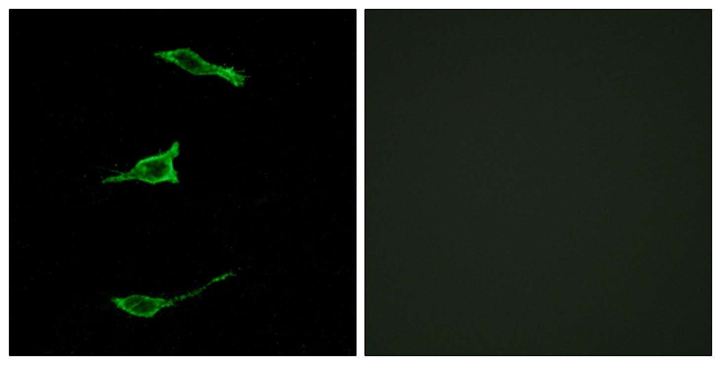
- Immunofluorescence analysis of LOVO cells, using FFAR1 Antibody. The picture on the right is blocked with the synthesized peptide.
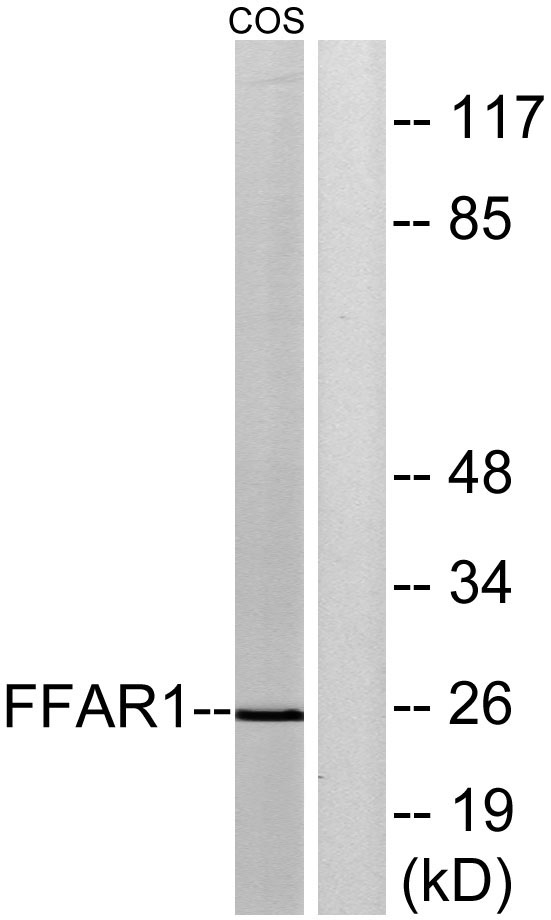
- Western blot analysis of lysates from COS7 cells, treated with forskolin 40nM 30', using FFAR1 Antibody. The lane on the right is blocked with the synthesized peptide.


