GluR-1 Polyclonal Antibody
- Catalog No.:YT1921
- Applications:WB;IHC;IF;ELISA
- Reactivity:Human;Mouse;Rat
- Target:
- GluR-1
- Fields:
- >>cAMP signaling pathway;>>Neuroactive ligand-receptor interaction;>>Circadian entrainment;>>Long-term potentiation;>>Retrograde endocannabinoid signaling;>>Glutamatergic synapse;>>Dopaminergic synapse;>>Long-term depression;>>Amyotrophic lateral sclerosis;>>Huntington disease;>>Spinocerebellar ataxia;>>Pathways of neurodegeneration - multiple diseases;>>Amphetamine addiction;>>Nicotine addiction
- Gene Name:
- GRIA1
- Protein Name:
- Glutamate receptor 1
- Human Gene Id:
- 2890
- Human Swiss Prot No:
- P42261
- Mouse Gene Id:
- 14799
- Mouse Swiss Prot No:
- P23818
- Rat Gene Id:
- 50592
- Rat Swiss Prot No:
- P19490
- Immunogen:
- The antiserum was produced against synthesized peptide derived from human GluR1. AA range:816-865
- Specificity:
- GluR-1 Polyclonal Antibody detects endogenous levels of GluR-1 protein.
- Formulation:
- Liquid in PBS containing 50% glycerol, 0.5% BSA and 0.02% sodium azide.
- Source:
- Polyclonal, Rabbit,IgG
- Dilution:
- WB 1:500 - 1:2000. IHC 1:100 - 1:300. IF 1:200 - 1:1000. ELISA: 1:40000. Not yet tested in other applications.
- Purification:
- The antibody was affinity-purified from rabbit antiserum by affinity-chromatography using epitope-specific immunogen.
- Concentration:
- 1 mg/ml
- Storage Stability:
- -15°C to -25°C/1 year(Do not lower than -25°C)
- Other Name:
- GRIA1;GLUH1;GLUR1;Glutamate receptor 1;GluR-1;AMPA-selective glutamate receptor 1;GluR-A;GluR-K1;Glutamate receptor ionotropic; AMPA 1;GluA1
- Observed Band(KD):
- 95kD
- Background:
- Glutamate receptors are the predominant excitatory neurotransmitter receptors in the mammalian brain and are activated in a variety of normal neurophysiologic processes. These receptors are heteromeric protein complexes with multiple subunits, each possessing transmembrane regions, and all arranged to form a ligand-gated ion channel. The classification of glutamate receptors is based on their activation by different pharmacologic agonists. This gene belongs to a family of alpha-amino-3-hydroxy-5-methyl-4-isoxazole propionate (AMPA) receptors. Alternatively spliced transcript variants encoding different isoforms have been found for this gene. [provided by RefSeq, Jul 2008],
- Function:
- function:Ionotropic glutamate receptor. L-glutamate acts as an excitatory neurotransmitter at many synapses in the central nervous system. Binding of the excitatory neurotransmitter L-glutamate induces a conformation change, leading to the opening of the cation channel, and thereby converts the chemical signal to an electrical impulse. The receptor then desensitizes rapidly and enters a transient inactive state, characterized by the presence of bound agonist.,miscellaneous:The postsynaptic actions of Glu are mediated by a variety of receptors that are named according to their selective agonists. This receptor binds AMPA (quisqualate) > glutamate > kainate.,PTM:Palmitoylated. Depalmitoylated upon glutamate stimulation. Cys-603 palmitoylation leads to Golgi retention and decreased cell surface expression. In contrast, Cys-829 palmitoylation does not affect cell surface expression but regul
- Subcellular Location:
- Cell membrane ; Multi-pass membrane protein . Endoplasmic reticulum membrane ; Multi-pass membrane protein . Cell junction, synapse, postsynaptic cell membrane ; Multi-pass membrane protein . Cell junction, synapse, postsynaptic density membrane ; Multi-pass membrane protein . Cell projection, dendrite . Cell projection, dendritic spine . Early endosome membrane ; Multi-pass membrane protein . Recycling endosome membrane ; Multi-pass membrane protein . Cell junction, synapse, presynapse . Cell junction, synapse . Interaction with CACNG2, CNIH2 and CNIH3 promotes cell surface expression. Colocalizes with PDLIM4 in early endosomes. Displays a somatodendritic localization and is excluded from axons in neurons (By similarity). Localized to cone photoreceptor pedicles (By similarity). .
- Expression:
- Widely expressed in brain.
- June 19-2018
- WESTERN IMMUNOBLOTTING PROTOCOL
- June 19-2018
- IMMUNOHISTOCHEMISTRY-PARAFFIN PROTOCOL
- June 19-2018
- IMMUNOFLUORESCENCE PROTOCOL
- September 08-2020
- FLOW-CYTOMEYRT-PROTOCOL
- May 20-2022
- Cell-Based ELISA│解您多样本WB检测之困扰
- July 13-2018
- CELL-BASED-ELISA-PROTOCOL-FOR-ACETYL-PROTEIN
- July 13-2018
- CELL-BASED-ELISA-PROTOCOL-FOR-PHOSPHO-PROTEIN
- July 13-2018
- Antibody-FAQs
- Products Images
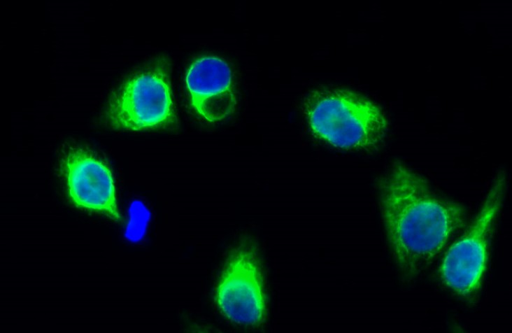
- Immunofluorescence analysis of Hela cell. 1,GluR-1 Polyclonal Antibody(green) was diluted at 1:200(4° overnight). 2, Goat Anti Rabbit Alexa Fluor 488 Catalog:RS3211 was diluted at 1:1000(room temperature, 50min). 3 DAPI(blue) 10min.
- Western Blot analysis of various cells using GluR-1 Polyclonal Antibody diluted at 1:500
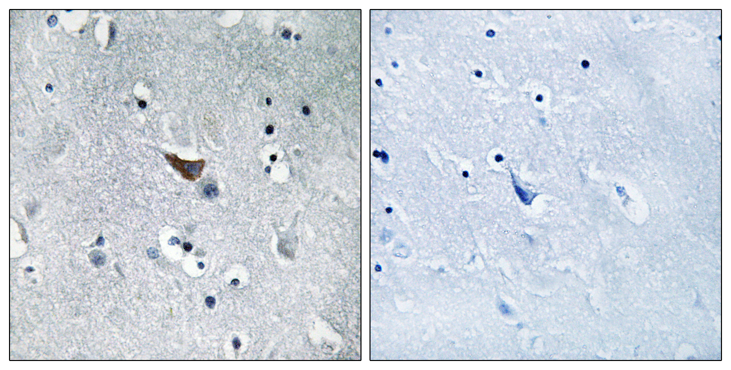
- Immunohistochemistry analysis of paraffin-embedded human brain tissue, using GluR1 Antibody. The picture on the right is blocked with the synthesized peptide.

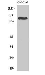
- Western blot analysis of lysates from COLO and Jurkat cells, using GluR1 Antibody. The lane on the right is blocked with the synthesized peptide.


