Galectin-9 Polyclonal Antibody
- Catalog No.:YT1841
- Applications:WB;IHC;IF;ELISA
- Reactivity:Human;Mouse;Rat
- Target:
- Galectin-9
- Gene Name:
- LGALS9
- Protein Name:
- Galectin-9
- Human Gene Id:
- 3965
- Human Swiss Prot No:
- O00182
- Mouse Gene Id:
- 16859
- Mouse Swiss Prot No:
- O08573
- Rat Gene Id:
- 25476
- Rat Swiss Prot No:
- P97840
- Immunogen:
- The antiserum was produced against synthesized peptide derived from human LEG9. AA range:51-100
- Specificity:
- Galectin-9 Polyclonal Antibody detects endogenous levels of Galectin-9 protein.
- Formulation:
- Liquid in PBS containing 50% glycerol, 0.5% BSA and 0.02% sodium azide.
- Source:
- Polyclonal, Rabbit,IgG
- Dilution:
- WB 1:500 - 1:2000. IHC 1:100 - 1:300. IF 1:200 - 1:1000. ELISA: 1:10000. Not yet tested in other applications.
- Purification:
- The antibody was affinity-purified from rabbit antiserum by affinity-chromatography using epitope-specific immunogen.
- Concentration:
- 1 mg/ml
- Storage Stability:
- -15°C to -25°C/1 year(Do not lower than -25°C)
- Other Name:
- LGALS9;Galectin-9;Gal-9;Ecalectin;Tumor antigen HOM-HD-21
- Observed Band(KD):
- 40kD
- Background:
- The galectins are a family of beta-galactoside-binding proteins implicated in modulating cell-cell and cell-matrix interactions. The protein encoded by this gene is an S-type lectin. It is overexpressed in Hodgkin's disease tissue and might participate in the interaction between the H&RS cells with their surrounding cells and might thus play a role in the pathogenesis of this disease and/or its associated immunodeficiency. Multiple alternatively spliced transcript variants have been found for this gene. [provided by RefSeq, Jul 2008],
- Function:
- alternative products:Additional isoforms seem to exist,domain:Contains two homologous but distinct carbohydrate-binding domains.,function:Binds galactosides. Has high affinity for the Forssman pentasaccharide. May play a role in thymocyte-epithelial interactions relevant to the biology of the thymus. Inhibits cell proliferation. The isoform Short acts as an eosinophil chemoattractant. Is a ligand for HAVCR2/TIM3. Induces T-helper type 1 lymphocyte (Th1) death.,online information:Galectin-9,similarity:Contains 2 galectin domains.,subcellular location:May also be secreted by a non-classical secretory pathway.,subunit:Monomer.,tissue specificity:Peripheral blood leukocytes and lymphatic tissues. Overexpressed in Hodgkin's disease tissue.,
- Subcellular Location:
- Cytoplasm . Nucleus . Secreted . May also be secreted by a non-classical secretory pathway (By similarity). Secreted by mesenchymal stromal cells upon IFNG stimulation (PubMed:23817958). .; [Isoform 2]: Secreted .; [Isoform 3]: Secreted .
- Expression:
- Peripheral blood leukocytes and lymphatic tissues. Expressed in lung, liver, breast and kidney with higher levels in tumor endothelial cells than normal endothelium (at protein level) (PubMed:24333696). Expressed in trophoblast cells in decidua and placenta in pregnancy (at protein level) (PubMed:23242525, PubMed:25578313). Isoform 2 is the most abundant isoform expressed in endothelial cells (PubMed:24333696). Upon endothelial cell activation isoform 2 expression decreases while expression of isoform 3 and isoform 5 increases (PubMed:24333696). Isoform 4 decreases in pathological pregnancy (PubMed:23242525).
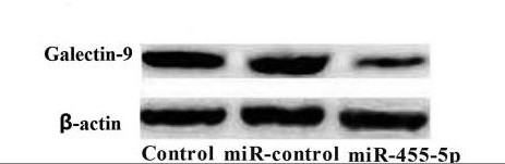
microRNA-22 downregulation of galectin-9 influences lymphocyte apoptosis and tumor cell proliferation in liver cancer. ONCOLOGY REPORTS 2015 Jul 31 WB Human Lo2 cells ,HepG2 cell, SMMC7721 cell
miR‑455‑5p functions as a potential oncogene by targeting galectin‑9 in colon cancer. Oncology Letters 2017 Jan 17 WB Human 1:1000 HT29 cell
程庆, 王晓华, and 杨斌. "寻常型银屑病患者体内半乳糖凝集素家族蛋白表达研究." 皮肤性病诊疗学杂志 27.3 (2020): 141-146.
- June 19-2018
- WESTERN IMMUNOBLOTTING PROTOCOL
- June 19-2018
- IMMUNOHISTOCHEMISTRY-PARAFFIN PROTOCOL
- June 19-2018
- IMMUNOFLUORESCENCE PROTOCOL
- September 08-2020
- FLOW-CYTOMEYRT-PROTOCOL
- May 20-2022
- Cell-Based ELISA│解您多样本WB检测之困扰
- July 13-2018
- CELL-BASED-ELISA-PROTOCOL-FOR-ACETYL-PROTEIN
- July 13-2018
- CELL-BASED-ELISA-PROTOCOL-FOR-PHOSPHO-PROTEIN
- July 13-2018
- Antibody-FAQs
- Products Images
- Yang, Qianqian, et al. "miR‑455‑5p functions as a potential oncogene by targeting galectin‑9 in colon cancer." Oncology letters 13.3 (2017): 1958-1964.
- Immunofluorescence analysis of Hela cell. 1,Galectin-9 Polyclonal Antibody(green) was diluted at 1:200(4° overnight). 2, Goat Anti Rabbit Alexa Fluor 488 Catalog:RS3211 was diluted at 1:1000(room temperature, 50min). 3 DAPI(blue) 10min.
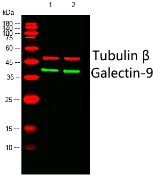
- Western blot analysis of lysates from 1) VEC, 2) HepG2 cells, (Green) primary antibody was diluted at 1:1000, 4°over night, Dylight 800 secondary antibody(Immunoway:RS23920)was diluted at 1:10000, 37° 1hour. (Red) Tubulin β Monoclonal Antibody(5G3) (Immunoway:YM3030) antibody was diluted at 1:5000 as loading control, 4° over night,Dylight 680 secondary antibody(Immunoway:RS23710)was diluted at 1:10000, 37° 1hour.
.jpg)
- Western Blot analysis of HepG2 cells using Galectin-9 Polyclonal Antibody diluted at 1:1000
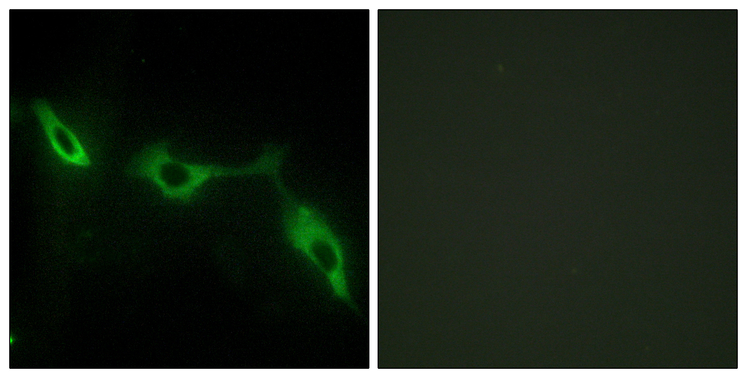
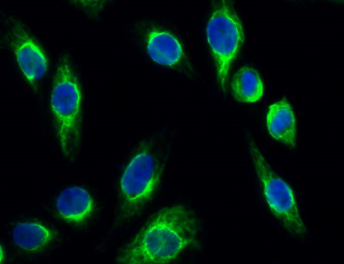
- Immunofluorescence analysis of NIH/3T3 cells, using LEG9 Antibody. The picture on the right is blocked with the synthesized peptide.
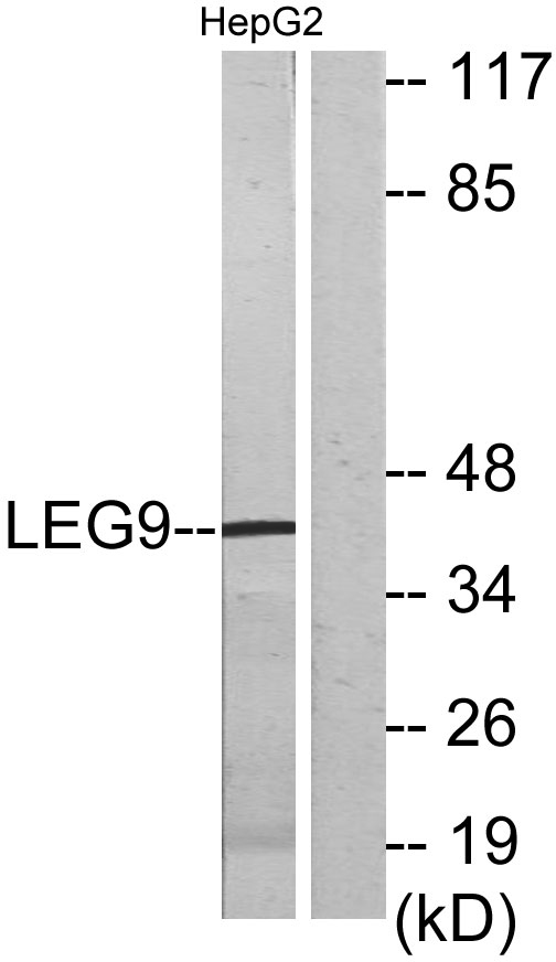
- Western blot analysis of lysates from HepG2 cells, using LEG9 Antibody. The lane on the right is blocked with the synthesized peptide.
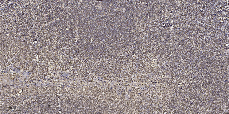
- Immunohistochemical analysis of paraffin-embedded human tonsil. 1, Antibody was diluted at 1:200(4° overnight). 2, Tris-EDTA,pH9.0 was used for antigen retrieval. 3,Secondary antibody was diluted at 1:200(room temperature, 45min).


