FOP Polyclonal Antibody
- Catalog No.:YT1736
- Applications:WB;IHC;IF;ELISA
- Reactivity:Human;Mouse;Rat
- Target:
- FOP
- Gene Name:
- FGFR1OP
- Protein Name:
- FGFR1 oncogene partner
- Human Gene Id:
- 11116
- Human Swiss Prot No:
- O95684
- Mouse Gene Id:
- 75296
- Mouse Swiss Prot No:
- Q66JX5
- Rat Gene Id:
- 683722
- Rat Swiss Prot No:
- Q4V7C1
- Immunogen:
- The antiserum was produced against synthesized peptide derived from human FGFR1 Oncogene Partner. AA range:341-390
- Specificity:
- FOP Polyclonal Antibody detects endogenous levels of FOP protein.
- Formulation:
- Liquid in PBS containing 50% glycerol, 0.5% BSA and 0.02% sodium azide.
- Source:
- Polyclonal, Rabbit,IgG
- Dilution:
- WB 1:500 - 1:2000. IHC 1:100 - 1:300. IF 1:200 - 1:1000. ELISA: 1:10000. Not yet tested in other applications.
- Purification:
- The antibody was affinity-purified from rabbit antiserum by affinity-chromatography using epitope-specific immunogen.
- Concentration:
- 1 mg/ml
- Storage Stability:
- -15°C to -25°C/1 year(Do not lower than -25°C)
- Other Name:
- FGFR1OP;FOP;FGFR1 oncogene partner
- Observed Band(KD):
- 43kD
- Background:
- FGFR1 oncogene partner(FGFR1OP) Homo sapiens This gene encodes a largely hydrophilic centrosomal protein that is required for anchoring microtubules to subcellular structures. A t(6;8)(q27;p11) chromosomal translocation, fusing this gene and the fibroblast growth factor receptor 1 (FGFR1) gene, has been found in cases of myeloproliferative disorder. The resulting chimeric protein contains the N-terminal leucine-rich region of this encoded protein fused to the catalytic domain of FGFR1. Alterations in this gene may also be associated with Crohn's disease, Graves' disease, and vitiligo. Alternatively spliced transcript variants that encode different proteins have been identified. [provided by RefSeq, Jul 2013],
- Function:
- disease:A chromosomal aberration involving FGFR1OP may be a cause of stem cell myeloproliferative disorder (MPD). Translocation t(6;8)(q27;p11) with FGFR1. MPD is characterized by myeloid hyperplasia, eosinophilia and T-cell or B-cell lymphoblastic lymphoma. In general it progresses to acute myeloid leukemia. The fusion proteins FGFR1OP-FGFR1 or FGFR1-FGFR1OP may exhibit constitutive kinase activity and be responsible for the transforming activity.,function:Required for anchoring microtubules to the centrosomes.,similarity:Contains 1 LisH domain.,subcellular location:Associated with gamma-tubulin.,subunit:Homodimer. Part of a ternary complex that contains CEP350, FGFR1OP and MAPRE1. Interacts directly with CEP350 and MAPRE1.,tissue specificity:Ubiquitous. Highly expressed in heart, liver, muscle, kidney, intestine, colon, adrenal gland, prostate, testis, and pancreas.,
- Subcellular Location:
- Cytoplasm, cytoskeleton, microtubule organizing center, centrosome . Cytoplasm, cytoskeleton, microtubule organizing center, centrosome, centriole . Cytoplasm, cytoskeleton, cilium basal body . Associated with gamma-tubulin (PubMed:16314388). Localizes on both mother and daughter centrioles (PubMed:28625565, PubMed:28428259). Localizes to an axial position on the mother centriole (PubMed:28625565). Localizes to the distal end of the centriole partly on the subdistal appendage region (PubMed:28659385). .
- Expression:
- Ubiquitous. Highly expressed in heart, liver, muscle, kidney, intestine, colon, adrenal gland, prostate, testis, and pancreas.
- June 19-2018
- WESTERN IMMUNOBLOTTING PROTOCOL
- June 19-2018
- IMMUNOHISTOCHEMISTRY-PARAFFIN PROTOCOL
- June 19-2018
- IMMUNOFLUORESCENCE PROTOCOL
- September 08-2020
- FLOW-CYTOMEYRT-PROTOCOL
- May 20-2022
- Cell-Based ELISA│解您多样本WB检测之困扰
- July 13-2018
- CELL-BASED-ELISA-PROTOCOL-FOR-ACETYL-PROTEIN
- July 13-2018
- CELL-BASED-ELISA-PROTOCOL-FOR-PHOSPHO-PROTEIN
- July 13-2018
- Antibody-FAQs
- Products Images
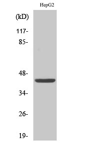
- Western Blot analysis of various cells using FOP Polyclonal Antibody
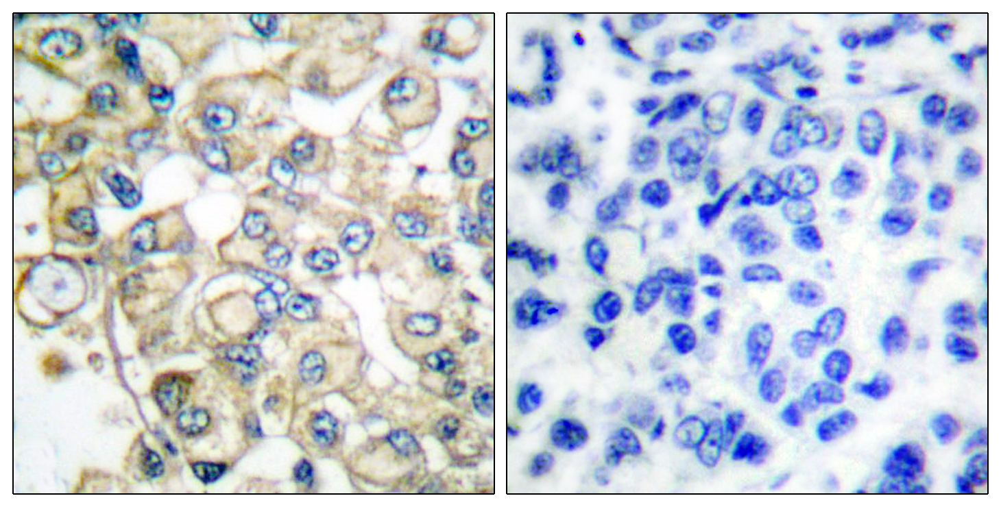
- Immunohistochemistry analysis of paraffin-embedded human breast carcinoma tissue, using FGFR1 Oncogene Partner Antibody. The picture on the right is blocked with the synthesized peptide.
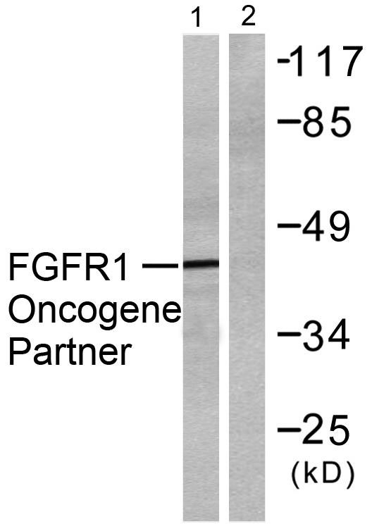
- Western blot analysis of lysates from HepG2 cells, using FGFR1 Oncogene Partner Antibody. The lane on the right is blocked with the synthesized peptide.


