ERK 8 Polyclonal Antibody
- Catalog No.:YT1630
- Applications:WB;IHC;IF;ELISA
- Reactivity:Human;Mouse
- Target:
- ERK8
- Fields:
- >>IL-17 signaling pathway
- Gene Name:
- MAPK15
- Protein Name:
- Mitogen-activated protein kinase 15
- Human Gene Id:
- 225689
- Human Swiss Prot No:
- Q8TD08
- Mouse Gene Id:
- 332110
- Mouse Swiss Prot No:
- Q80Y86
- Immunogen:
- The antiserum was produced against synthesized peptide derived from human MAPK15. AA range:141-190
- Specificity:
- ERK 8 Polyclonal Antibody detects endogenous levels of ERK 8 protein.
- Formulation:
- Liquid in PBS containing 50% glycerol, 0.5% BSA and 0.02% sodium azide.
- Source:
- Polyclonal, Rabbit,IgG
- Dilution:
- WB 1:500 - 1:2000. IHC 1:100 - 1:300. IF 1:200 - 1:1000. ELISA: 1:10000. Not yet tested in other applications.
- Purification:
- The antibody was affinity-purified from rabbit antiserum by affinity-chromatography using epitope-specific immunogen.
- Concentration:
- 1 mg/ml
- Storage Stability:
- -15°C to -25°C/1 year(Do not lower than -25°C)
- Other Name:
- MAPK15;ERK7;ERK8;Mitogen-activated protein kinase 15;MAP kinase 15;MAPK 15;Extracellular signal-regulated kinase 7;ERK-7;Extracellular signal-regulated kinase 8;ERK-8
- Observed Band(KD):
- 60kD
- Background:
- catalytic activity:ATP + a protein = ADP + a phosphoprotein.,domain:The N-terminal region (1-20) is the minimal region necessary for ubiquitination and further proteosomal degradation.,domain:The TXY motif contains the threonine and tyrosine residues whose phosphorylation activates the MAP kinases.,enzyme regulation:Activated by threonine and tyrosine phosphorylation. Inhibited by dual specificity phosphatases, such as DUSP1.,function:In vitro, phosphorylates MBP.,PTM:Dually phosphorylated on Thr-175 and Tyr-177, which activates the enzyme. Autophosphorylated on threonine and tyrosine residues in vitro.,PTM:Ubiquitinated. Ubiquitination may allow its tight kinase activity regulation and rapid turnover. May be ubiquitinated by a SCF E3 ligase.,similarity:Belongs to the protein kinase superfamily. CMGC Ser/Thr protein kinase family. MAP kinase subfamily.,similarity:Contains 1 protein kinase domain.,subunit:Interacts with CSK/c-Src, ABL1, RET and TGFB1I1.,tissue specificity:Widely expressed with a maximal expression in lung and kidney.,
- Function:
- catalytic activity:ATP + a protein = ADP + a phosphoprotein.,domain:The N-terminal region (1-20) is the minimal region necessary for ubiquitination and further proteosomal degradation.,domain:The TXY motif contains the threonine and tyrosine residues whose phosphorylation activates the MAP kinases.,enzyme regulation:Activated by threonine and tyrosine phosphorylation. Inhibited by dual specificity phosphatases, such as DUSP1.,function:In vitro, phosphorylates MBP.,PTM:Dually phosphorylated on Thr-175 and Tyr-177, which activates the enzyme. Autophosphorylated on threonine and tyrosine residues in vitro.,PTM:Ubiquitinated. Ubiquitination may allow its tight kinase activity regulation and rapid turnover. May be ubiquitinated by a SCF E3 ligase.,similarity:Belongs to the protein kinase superfamily. CMGC Ser/Thr protein kinase family. MAP kinase subfamily.,similarity:Contains 1 protein kinas
- Subcellular Location:
- Cytoplasm, cytoskeleton, cilium basal body . Cell junction, tight junction . Cytoplasm, cytoskeleton, microtubule organizing center, centrosome, centriole . Cytoplasmic vesicle, autophagosome . Golgi apparatus . Nucleus . Cytoplasm . Cytoplasm, cytoskeleton, spindle . Co-localizes to the cytoplasm only in presence of ESRRA (PubMed:21190936). Translocates to the nucleus upon activation (PubMed:20638370). At prometaphase I, metaphase I (MI), anaphase I, telophase I, and metaphase II (MII) stages, is stably detected at the spindle (By similarity). .
- Expression:
- Widely expressed with a maximal expression in lung and kidney.
- June 19-2018
- WESTERN IMMUNOBLOTTING PROTOCOL
- June 19-2018
- IMMUNOHISTOCHEMISTRY-PARAFFIN PROTOCOL
- June 19-2018
- IMMUNOFLUORESCENCE PROTOCOL
- September 08-2020
- FLOW-CYTOMEYRT-PROTOCOL
- May 20-2022
- Cell-Based ELISA│解您多样本WB检测之困扰
- July 13-2018
- CELL-BASED-ELISA-PROTOCOL-FOR-ACETYL-PROTEIN
- July 13-2018
- CELL-BASED-ELISA-PROTOCOL-FOR-PHOSPHO-PROTEIN
- July 13-2018
- Antibody-FAQs
- Products Images
- Western Blot analysis of various cells using ERK 8 Polyclonal Antibody cells nucleus extracted by Minute TM Cytoplasmic and Nuclear Fractionation kit (SC-003,Inventbiotech,MN,USA).
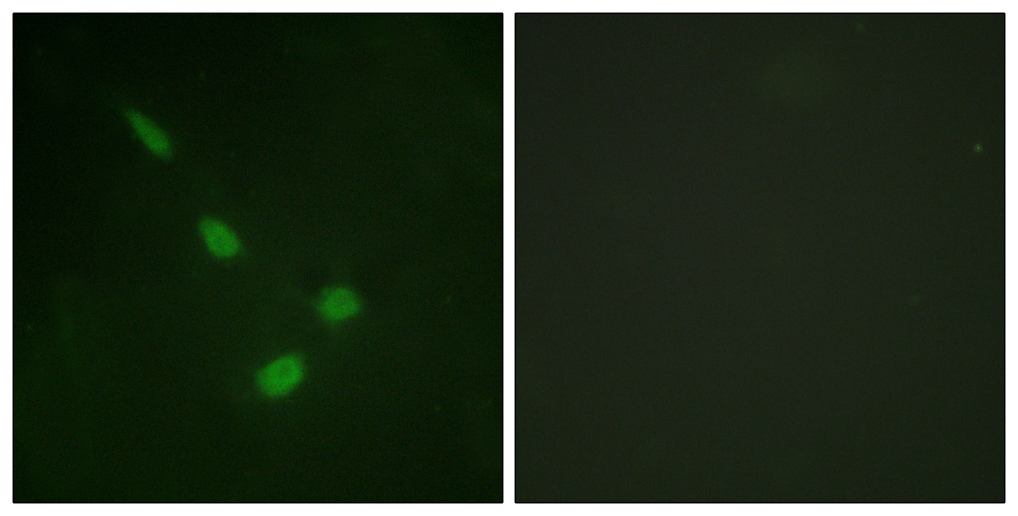
- Immunofluorescence analysis of NIH/3T3 cells, using ERK8 Antibody. The picture on the right is blocked with the synthesized peptide.
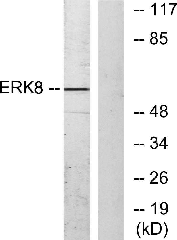
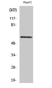
- Western blot analysis of lysates from HepG2 cells, using ERK8 Antibody. The lane on the right is blocked with the synthesized peptide.
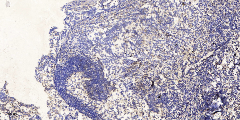
- Immunohistochemical analysis of paraffin-embedded human tonsil. 1, Antibody was diluted at 1:200(4° overnight). 2, Tris-EDTA,pH9.0 was used for antigen retrieval. 3,Secondary antibody was diluted at 1:200(room temperature, 30min).


