EphB6 Polyclonal Antibody
- Catalog No.:YT1588
- Applications:WB;IHC;IF;ELISA
- Reactivity:Human;Mouse;Rat
- Target:
- EphB6
- Fields:
- >>Axon guidance
- Gene Name:
- EPHB6
- Protein Name:
- Ephrin type-B receptor 6
- Human Gene Id:
- 2051
- Human Swiss Prot No:
- O15197
- Mouse Gene Id:
- 13848
- Mouse Swiss Prot No:
- O08644
- Rat Gene Id:
- 312275
- Rat Swiss Prot No:
- P0C0K7
- Immunogen:
- The antiserum was produced against synthesized peptide derived from human EPHB6. AA range:861-910
- Specificity:
- EphB6 Polyclonal Antibody detects endogenous levels of EphB6 protein.
- Formulation:
- Liquid in PBS containing 50% glycerol, 0.5% BSA and 0.02% sodium azide.
- Source:
- Polyclonal, Rabbit,IgG
- Dilution:
- WB 1:500 - 1:2000. IHC 1:100 - 1:300. IF 1:200 - 1:1000. ELISA: 1:5000. Not yet tested in other applications.
- Purification:
- The antibody was affinity-purified from rabbit antiserum by affinity-chromatography using epitope-specific immunogen.
- Concentration:
- 1 mg/ml
- Storage Stability:
- -15°C to -25°C/1 year(Do not lower than -25°C)
- Other Name:
- EPHB6;Ephrin type-B receptor 6;HEP;Tyrosine-protein kinase-defective receptor EPH-6
- Observed Band(KD):
- 119kD
- Background:
- This gene encodes a member of a family of transmembrane proteins that function as receptors for ephrin-B family proteins. Unlike other members of this family, the encoded protein does not contain a functional kinase domain. Activity of this protein can influence cell adhesion and migration. Expression of this gene is downregulated during tumor progression, suggesting that the protein may suppress tumor invasion and metastasis. Alternative splicing results in multiple transcript variants. [provided by RefSeq, Jul 2013],
- Function:
- domain:The protein kinase domain is predicted to be catalytically inactive. Its extracellular domain is capable of promoting cell adhesion and migration in response to low concentrations of ephrin-B2, but its cytoplasmic domain is essential for cell repulsion and inhibition of migration induced by high concentrations of ephrin-B2.,function:Kinase-defective receptor for members of the ephrin-B family. Binds to ephrin-B1 and ephrin-B2. Modulates cell adhesion and migration by exerting both positive and negative effects upon stimulation with ephrin-B2. Inhibits JNK activation, T cell receptor-induced IL-2 secretion and CD25 expression upon stimulation with ephrin-B2.,PTM:Ligand-binding increases phosphorylation on tyrosine residues. Phosphorylation on tyrosine residues is mediated by transphosphorylation by the catalytically active EPHB1 in a ligand-independent manner. Tyrosine phosphorylat
- Subcellular Location:
- Membrane; Single-pass type I membrane protein.; [Isoform 3]: Secreted .
- Expression:
- Expressed in brain. Expressed in non invasive breast carcinoma cell lines (at protein level). Strong expression in brain and pancreas, and weak expression in other tissues, such as heart, placenta, lung, liver, skeletal muscle and kidney. Expressed in breast non invasive tumors but not in metastatic lesions. Isoform 3 is expressed in cell lines of glioblastomas, anaplastic astrocytomas, gliosarcomas and astrocytomas. Isoform 3 is not detected in normal tissues.
- June 19-2018
- WESTERN IMMUNOBLOTTING PROTOCOL
- June 19-2018
- IMMUNOHISTOCHEMISTRY-PARAFFIN PROTOCOL
- June 19-2018
- IMMUNOFLUORESCENCE PROTOCOL
- September 08-2020
- FLOW-CYTOMEYRT-PROTOCOL
- May 20-2022
- Cell-Based ELISA│解您多样本WB检测之困扰
- July 13-2018
- CELL-BASED-ELISA-PROTOCOL-FOR-ACETYL-PROTEIN
- July 13-2018
- CELL-BASED-ELISA-PROTOCOL-FOR-PHOSPHO-PROTEIN
- July 13-2018
- Antibody-FAQs
- Products Images
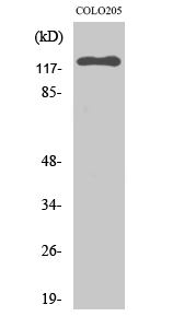
- Western Blot analysis of various cells using EphB6 Polyclonal Antibody
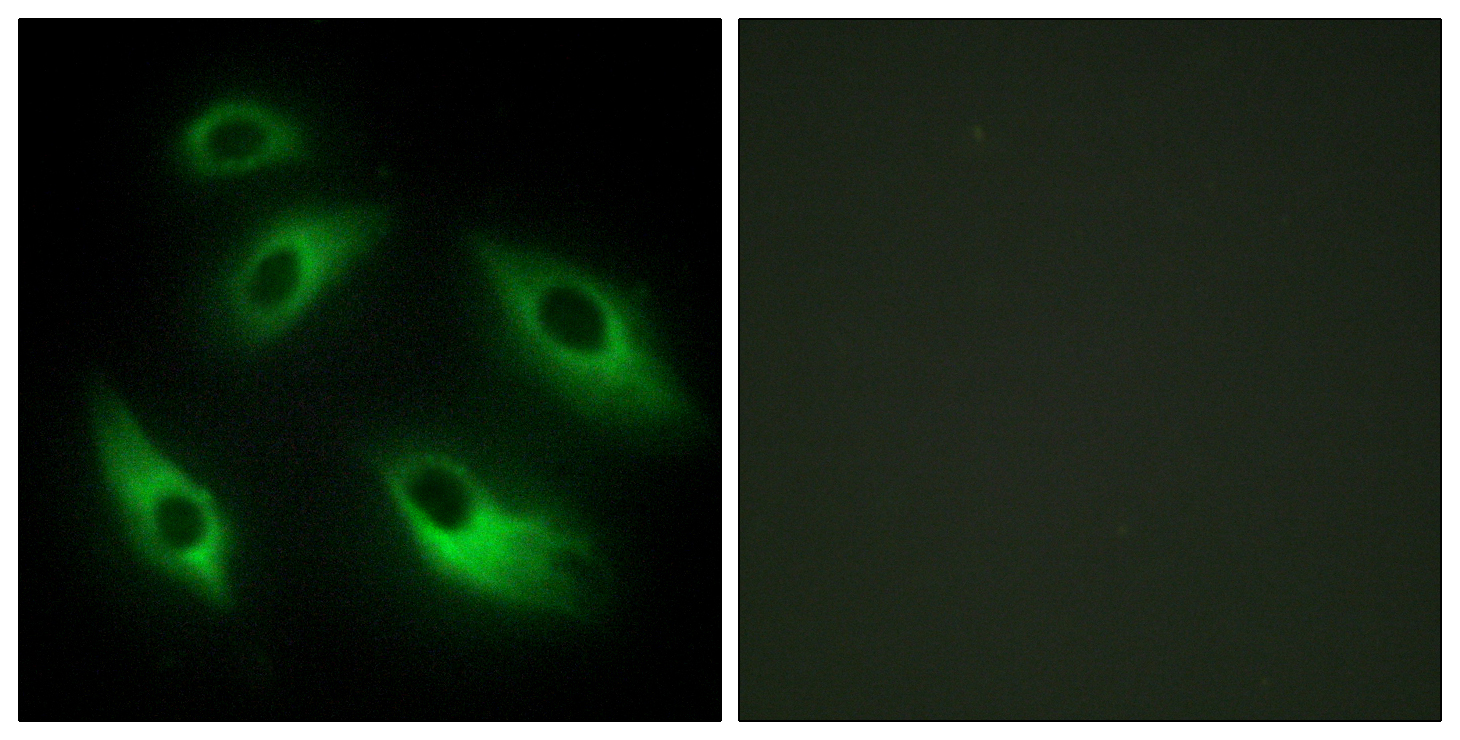
- Immunofluorescence analysis of HeLa cells, using EPHB6 Antibody. The picture on the right is blocked with the synthesized peptide.
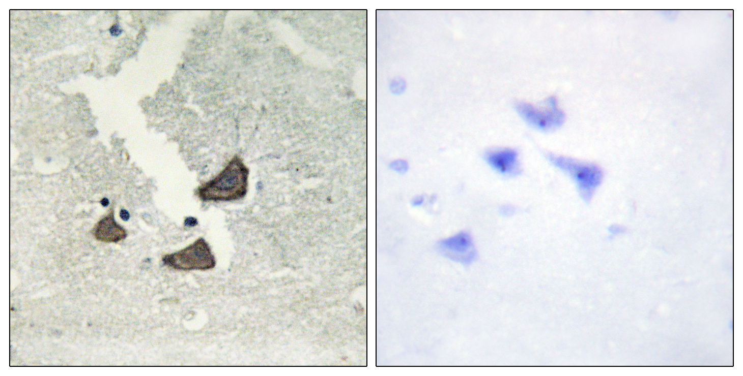
- Immunohistochemistry analysis of paraffin-embedded human brain tissue, using EPHB6 Antibody. The picture on the right is blocked with the synthesized peptide.
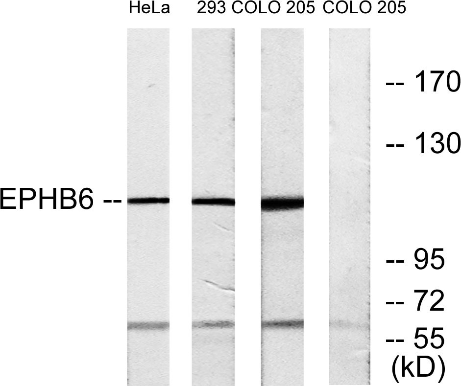
- Western blot analysis of lysates from COLO, 293, and HeLa cells, using EPHB6 Antibody. The lane on the right is blocked with the synthesized peptide.


