Endophilin I Polyclonal Antibody
- Catalog No.:YT1559
- Applications:WB;ELISA
- Reactivity:Human;Mouse;Rat
- Target:
- Endophilin I
- Fields:
- >>Endocytosis
- Gene Name:
- SH3GL2
- Protein Name:
- Endophilin-A1
- Human Gene Id:
- 6456
- Human Swiss Prot No:
- Q99962
- Mouse Gene Id:
- 20404
- Mouse Swiss Prot No:
- Q62420
- Rat Gene Id:
- 116743
- Rat Swiss Prot No:
- O35179
- Immunogen:
- Synthesized peptide derived from Endophilin I . at AA range: 30-110
- Specificity:
- Endophilin I Polyclonal Antibody detects endogenous levels of Endophilin I protein.
- Formulation:
- Liquid in PBS containing 50% glycerol, 0.5% BSA and 0.02% sodium azide.
- Source:
- Polyclonal, Rabbit,IgG
- Dilution:
- WB 1:500 - 1:2000. ELISA: 1:10000. Not yet tested in other applications.
- Purification:
- The antibody was affinity-purified from rabbit antiserum by affinity-chromatography using epitope-specific immunogen.
- Concentration:
- 1 mg/ml
- Storage Stability:
- -15°C to -25°C/1 year(Do not lower than -25°C)
- Other Name:
- SH3GL2;CNSA2;SH3D2A;Endophilin-A1;EEN-B1;Endophilin-1;SH3 domain protein 2A;SH3 domain-containing GRB2-like protein 2
- Observed Band(KD):
- 39kD
- Background:
- domain:An N-terminal amphipathic helix, the BAR domain and a second amphipathic helix inserted into helix 1 of the BAR domain (N-BAR domain) induce membrane curvature and bind curved membranes. The BAR domain dimer forms a rigid crescent shaped bundle of helices with the pair of second amphipathic helices protruding towards the membrane-binding surface.,function:Implicated in synaptic vesicle endocytosis. May recruit other proteins to membranes with high curvature.,miscellaneous:HeLa cells expressing the N-BAR domain of SH3GL2 show tubulation of the plasma membrane. The N-BAR domain binds liposomes and induces formation of tubules from liposomes. The N-terminal amphipathic helix is required for liposome binding. The second amphipathic helix enhances liposome tubulation.,similarity:Belongs to the endophilin family.,similarity:Contains 1 BAR domain.,similarity:Contains 1 SH3 domain.,subcellular location:Concentrated in presynaptic nerve terminals in neurons.,subunit:Monomer; in cytoplasm. Homodimer; when associated with membranes (By similarity). Interacts with SYNJ1 and DNM1. Interacts with MAP4K3; the interaction appears to regulate MAP4K3-mediated JNK activation. Interacts with PDCD6IP.,tissue specificity:Brain, mostly in frontal cortex. Expressed at high level in fetal cerebellum.,
- Function:
- domain:An N-terminal amphipathic helix, the BAR domain and a second amphipathic helix inserted into helix 1 of the BAR domain (N-BAR domain) induce membrane curvature and bind curved membranes. The BAR domain dimer forms a rigid crescent shaped bundle of helices with the pair of second amphipathic helices protruding towards the membrane-binding surface.,function:Implicated in synaptic vesicle endocytosis. May recruit other proteins to membranes with high curvature.,miscellaneous:HeLa cells expressing the N-BAR domain of SH3GL2 show tubulation of the plasma membrane. The N-BAR domain binds liposomes and induces formation of tubules from liposomes. The N-terminal amphipathic helix is required for liposome binding. The second amphipathic helix enhances liposome tubulation.,similarity:Belongs to the endophilin family.,similarity:Contains 1 BAR domain.,similarity:Contains 1 SH3 domain.,subcel
- Subcellular Location:
- Cytoplasm . Membrane ; Peripheral membrane protein . Early endosome . Cell junction, synapse, presynapse .
- Expression:
- Brain, mostly in frontal cortex. Expressed at high level in fetal cerebellum.
- June 19-2018
- WESTERN IMMUNOBLOTTING PROTOCOL
- June 19-2018
- IMMUNOHISTOCHEMISTRY-PARAFFIN PROTOCOL
- June 19-2018
- IMMUNOFLUORESCENCE PROTOCOL
- September 08-2020
- FLOW-CYTOMEYRT-PROTOCOL
- May 20-2022
- Cell-Based ELISA│解您多样本WB检测之困扰
- July 13-2018
- CELL-BASED-ELISA-PROTOCOL-FOR-ACETYL-PROTEIN
- July 13-2018
- CELL-BASED-ELISA-PROTOCOL-FOR-PHOSPHO-PROTEIN
- July 13-2018
- Antibody-FAQs
- Products Images
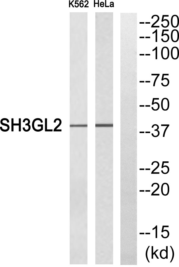
- Western blot analysis of SH3GL2 Antibody. The lane on the right is blocked with the SH3GL2 peptide.


