EMR3 Polyclonal Antibody
- Catalog No.:YT1547
- Applications:WB;IF;ELISA
- Reactivity:Human
- Target:
- EMR3
- Gene Name:
- EMR3
- Protein Name:
- EGF-like module-containing mucin-like hormone receptor-like 3
- Human Gene Id:
- 84658
- Human Swiss Prot No:
- Q9BY15
- Immunogen:
- The antiserum was produced against synthesized peptide derived from human EMR3. AA range:603-652
- Specificity:
- EMR3 Polyclonal Antibody detects endogenous levels of EMR3 protein.
- Formulation:
- Liquid in PBS containing 50% glycerol, 0.5% BSA and 0.02% sodium azide.
- Source:
- Polyclonal, Rabbit,IgG
- Dilution:
- WB 1:500 - 1:2000. IF 1:200 - 1:1000. ELISA: 1:10000. Not yet tested in other applications.
- Purification:
- The antibody was affinity-purified from rabbit antiserum by affinity-chromatography using epitope-specific immunogen.
- Concentration:
- 1 mg/ml
- Storage Stability:
- -15°C to -25°C/1 year(Do not lower than -25°C)
- Other Name:
- EMR3;EGF-like module-containing mucin-like hormone receptor-like 3;EGF-like module receptor 3
- Observed Band(KD):
- 70kD
- Background:
- This gene encodes a member of the class B seven-span transmembrane (TM7) receptor family expressed predominantly by cells of the immune system. Family members are characterized by an extended extracellular region with a variable number of N-terminal epidermal growth factor (EGF)-like domains coupled to a TM7 domain via a mucin-like spacer domain. This gene is closely linked to the gene encoding egf-like molecule containing mucin-like hormone receptor 2 on chromosome 19. This protein may play a role in myeloid-myeloid interactions during immune and inflammatory responses. Alternative splicing results in multiple transcript variants encoding different isoforms. [provided by RefSeq, Jan 2014],
- Function:
- function:Receptor probably involved in myeloid interactions during immune and inflammatory responses. A ligand for the soluble form of this receptor is present at the surface of momocytes-derived macrophages and activated neutrophils.,PTM:Proteolytically cleaved into 2 subunits, an extracellular alpha subunit and a seven-transmembrane subunit.,similarity:Belongs to the G-protein coupled receptor 2 family. LN-TM7 subfamily.,similarity:Contains 1 GPS domain.,similarity:Contains 2 EGF-like domains.,subcellular location:A soluble form is also detected (alpha subunit).,subunit:Forms a heterodimer, consisting of a large extracellular region (alpha subunit) non-covalently linked to a seven-transmembrane moiety (beta subunit).,tissue specificity:Displays a predominantly leukocyte-restricted expression, with highest levels in neutrophils, monocytes and macrophages.,
- Subcellular Location:
- Cell membrane; Multi-pass membrane protein .; [Isoform 3]: Secreted.
- Expression:
- Displays a predominantly leukocyte-restricted expression, with highest levels in neutrophils, monocytes and macrophages.
- June 19-2018
- WESTERN IMMUNOBLOTTING PROTOCOL
- June 19-2018
- IMMUNOHISTOCHEMISTRY-PARAFFIN PROTOCOL
- June 19-2018
- IMMUNOFLUORESCENCE PROTOCOL
- September 08-2020
- FLOW-CYTOMEYRT-PROTOCOL
- May 20-2022
- Cell-Based ELISA│解您多样本WB检测之困扰
- July 13-2018
- CELL-BASED-ELISA-PROTOCOL-FOR-ACETYL-PROTEIN
- July 13-2018
- CELL-BASED-ELISA-PROTOCOL-FOR-PHOSPHO-PROTEIN
- July 13-2018
- Antibody-FAQs
- Products Images
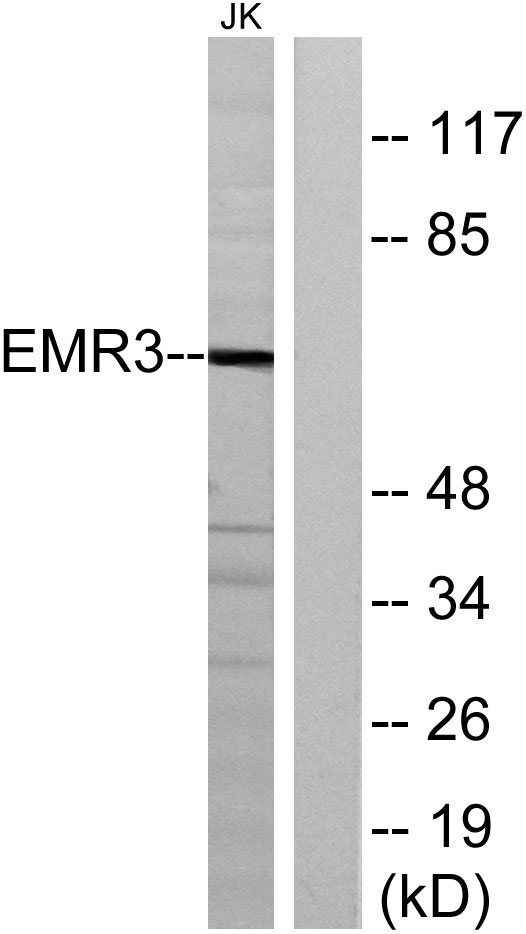
- Western blot analysis of lysates from Jurkat cells, using EMR3 Antibody. The lane on the right is blocked with the synthesized peptide.


