EMR2 Polyclonal Antibody
- Catalog No.:YT1546
- Applications:WB;IF;ELISA
- Reactivity:Human
- Target:
- EMR2
- Gene Name:
- EMR2
- Protein Name:
- EGF-like module-containing mucin-like hormone receptor-like 2
- Human Gene Id:
- 30817
- Human Swiss Prot No:
- Q9UHX3
- Immunogen:
- The antiserum was produced against synthesized peptide derived from human EMR2. AA range:765-814
- Specificity:
- EMR2 Polyclonal Antibody detects endogenous levels of EMR2 protein.
- Formulation:
- Liquid in PBS containing 50% glycerol, 0.5% BSA and 0.02% sodium azide.
- Source:
- Polyclonal, Rabbit,IgG
- Dilution:
- WB 1:500 - 1:2000. IF 1:200 - 1:1000. ELISA: 1:20000. Not yet tested in other applications.
- Purification:
- The antibody was affinity-purified from rabbit antiserum by affinity-chromatography using epitope-specific immunogen.
- Concentration:
- 1 mg/ml
- Storage Stability:
- -15°C to -25°C/1 year(Do not lower than -25°C)
- Other Name:
- EMR2;EGF-like module-containing mucin-like hormone receptor-like 2;EGF-like module receptor 2;CD antigen CD312
- Observed Band(KD):
- 85kD
- Background:
- This gene encodes a member of the class B seven-span transmembrane (TM7) subfamily of G-protein coupled receptors. These proteins are characterized by an extended extracellular region with a variable number of N-terminal epidermal growth factor-like domains coupled to a TM7 domain via a mucin-like spacer domain. The encoded protein is expressed mainly in myeloid cells where it promotes cell-cell adhesion through interaction with chondroitin sulfate chains. This gene is situated in a cluster of related genes on chromosome 19. Alternatively spliced transcript variants encoding multiple isoforms have been observed for this gene. [provided by RefSeq, Aug 2012],
- Function:
- alternative products:A number of isoforms are produced. A number of isoforms consisting of various number of EGF-like domains seems to exist. A soluble form due to a frameshift which introduced a stop codon immediately before the first TM domain is also detected,domain:Binding to chondroitin sulfate is mediated by the fourth EGF domain.,domain:The GPS domain is necessary, but not sufficient for receptor cleavage, which require the entire extracellular stalk.,function:Receptor probably involved in cell attachment.,PTM:Proteolytically cleaved into 2 subunits, an extracellular alpha subunit and a seven-transmembrane subunit.,similarity:Belongs to the G-protein coupled receptor 2 family. LN-TM7 subfamily.,similarity:Contains 1 GPS domain.,similarity:Contains 5 EGF-like domains.,subunit:Forms a heterodimer, consisting of a large extracellular region non-covalently linked to a seven-transmembr
- Subcellular Location:
- Cell membrane ; Multi-pass membrane protein. Cell projection, ruffle membrane ; Multi-pass membrane protein. Localized at the leading edge of migrating cells. .
- Expression:
- Expression is restricted to myeloid cells. Highest expression was found in peripheral blood leukocytes, followed by spleen and lymph nodes, with intermediate to low levels in thymus, bone marrow, fetal liver, placenta, and lung, and no expression in heart, brain, skeletal muscle, kidney, or pancreas. Expression is also detected in monocyte/macrophage and Jurkat cell lines but not in other cell lines tested. High expression in mast cells (PubMed:26841242).
- June 19-2018
- WESTERN IMMUNOBLOTTING PROTOCOL
- June 19-2018
- IMMUNOHISTOCHEMISTRY-PARAFFIN PROTOCOL
- June 19-2018
- IMMUNOFLUORESCENCE PROTOCOL
- September 08-2020
- FLOW-CYTOMEYRT-PROTOCOL
- May 20-2022
- Cell-Based ELISA│解您多样本WB检测之困扰
- July 13-2018
- CELL-BASED-ELISA-PROTOCOL-FOR-ACETYL-PROTEIN
- July 13-2018
- CELL-BASED-ELISA-PROTOCOL-FOR-PHOSPHO-PROTEIN
- July 13-2018
- Antibody-FAQs
- Products Images
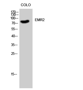
- Western Blot analysis of COLO cells using EMR2 Polyclonal Antibody
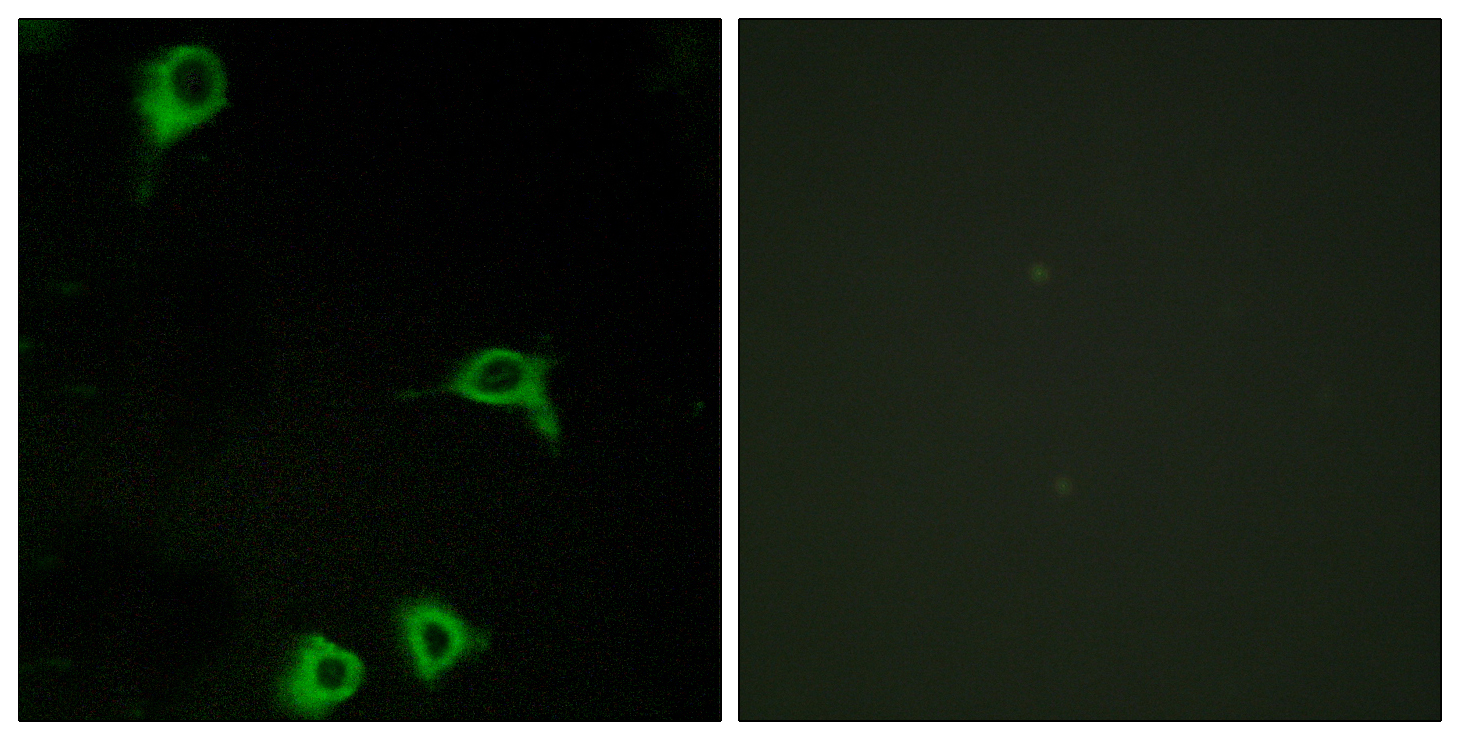
- Immunofluorescence analysis of COS7 cells, using EMR2 Antibody. The picture on the right is blocked with the synthesized peptide.
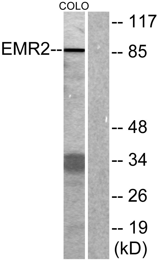
- Western blot analysis of lysates from COLO cells, using EMR2 Antibody. The lane on the right is blocked with the synthesized peptide.
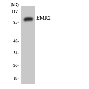
- Western blot analysis of the lysates from HUVECcells using EMR2 antibody.


