Emp Polyclonal Antibody
- Catalog No.:YT1543
- Applications:WB;IHC;IF;ELISA
- Reactivity:Human;Mouse;Rat
- Target:
- Emp
- Gene Name:
- MAEA
- Protein Name:
- Macrophage erythroblast attacher
- Human Gene Id:
- 10296
- Human Swiss Prot No:
- Q7L5Y9
- Mouse Gene Id:
- 59003
- Mouse Swiss Prot No:
- Q4VC33
- Rat Gene Id:
- 298982
- Rat Swiss Prot No:
- Q5RKJ1
- Immunogen:
- The antiserum was produced against synthesized peptide derived from human MAEA. AA range:181-230
- Specificity:
- Emp Polyclonal Antibody detects endogenous levels of Emp protein.
- Formulation:
- Liquid in PBS containing 50% glycerol, 0.5% BSA and 0.02% sodium azide.
- Source:
- Polyclonal, Rabbit,IgG
- Dilution:
- WB 1:500 - 1:2000. IHC 1:100 - 1:300. ELISA: 1:20000.. IF 1:50-200
- Purification:
- The antibody was affinity-purified from rabbit antiserum by affinity-chromatography using epitope-specific immunogen.
- Concentration:
- 1 mg/ml
- Storage Stability:
- -15°C to -25°C/1 year(Do not lower than -25°C)
- Other Name:
- MAEA;EMP;HLC10;PIG5;Macrophage erythroblast attacher;Cell proliferation-inducing gene 5 protein;Erythroblast macrophage protein;Human lung cancer oncogene 10 protein;HLC-10
- Observed Band(KD):
- 45kD
- Background:
- This gene encodes a protein that mediates the attachment of erythroblasts to macrophages. This attachment promotes terminal maturation and enucleation of erythroblasts, presumably by suppressing apoptosis. The encoded protein is an integral membrane protein with the N-terminus on the extracellular side and the C-terminus on the cytoplasmic side of the cell. Alternative splicing results in multiple transcript variants. [provided by RefSeq, Jul 2014],
- Function:
- developmental stage:Localized with condensed chromatin at prophase; Detected in nuclear spindle poles at metaphase and in the contractile ring during telophase and cytokinesis.,function:Play a role in erythroblast enucleation and in the development of the mature macrophages. Mediates the attachment of erythroid cell to mature macrophages, in correlation with the presence of MAEA at cell surface of mature macrophages; This MAEA-mediated contact inhibits erythroid cells apoptosis. Participates to erythroblastic island formation, which is the functional unit of definitive erythropoiesis. Associates with F-actin to regulate actin distribution in erythroblasts and macrophages. May contribute to nuclear architecture and cells division events.,similarity:Contains 1 CTLH domain.,similarity:Contains 1 LisH domain.,subcellular location:Localized as nuclear speckled-like pattern.,subunit:Form a com
- Subcellular Location:
- Cytoplasm . Nucleus, nucleoplasm . Nucleus matrix . Cell membrane . Cytoplasm, cytoskeleton . Detected in a nuclear, speckled-like pattern (PubMed:16510120). Localized with condensed chromatin at prophase; Detected in nuclear spindle poles at metaphase and in the contractile ring during telophase and cytokinesis (PubMed:16510120). Present in cytoplasm, nuclear matrix and at the cell surface in macrophages; predominantly nuclear in immature macrophages and predominantly detected at the cell surface in mature macrophages. Colocalizes with F-actin in macrophages (By similarity). .
- Expression:
- Detected at macrophage membranes (at protein level). Ubiquitous.
- June 19-2018
- WESTERN IMMUNOBLOTTING PROTOCOL
- June 19-2018
- IMMUNOHISTOCHEMISTRY-PARAFFIN PROTOCOL
- June 19-2018
- IMMUNOFLUORESCENCE PROTOCOL
- September 08-2020
- FLOW-CYTOMEYRT-PROTOCOL
- May 20-2022
- Cell-Based ELISA│解您多样本WB检测之困扰
- July 13-2018
- CELL-BASED-ELISA-PROTOCOL-FOR-ACETYL-PROTEIN
- July 13-2018
- CELL-BASED-ELISA-PROTOCOL-FOR-PHOSPHO-PROTEIN
- July 13-2018
- Antibody-FAQs
- Products Images
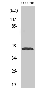
- Western Blot analysis of various cells using Emp Polyclonal Antibody diluted at 1:500
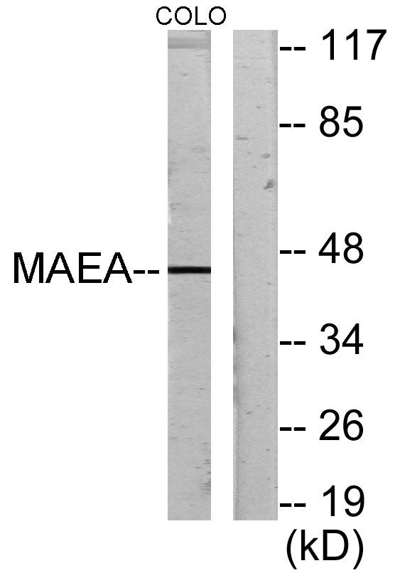
- Western blot analysis of lysates from COLO205 cells, using MAEA Antibody. The lane on the right is blocked with the synthesized peptide.
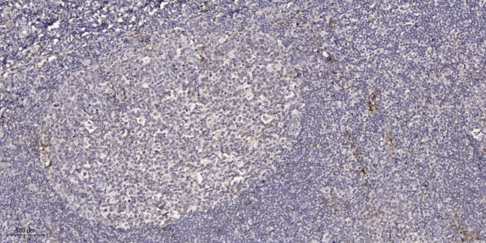
- Immunohistochemical analysis of paraffin-embedded human tonsil. 1, Antibody was diluted at 1:200(4° overnight). 2, Tris-EDTA,pH9.0 was used for antigen retrieval. 3,Secondary antibody was diluted at 1:200(room temperature, 45min).


