EMMPRIN Polyclonal Antibody
- Catalog No.:YT1542
- Applications:WB;IHC;IF;ELISA
- Reactivity:Human;Mouse;Rat
- Target:
- EMMPRIN
- Gene Name:
- BSG
- Protein Name:
- Basigin
- Human Gene Id:
- 682
- Human Swiss Prot No:
- P35613
- Mouse Gene Id:
- 12215
- Mouse Swiss Prot No:
- P18572
- Rat Gene Id:
- 25246
- Rat Swiss Prot No:
- P26453
- Immunogen:
- The antiserum was produced against synthesized peptide derived from human CD147. AA range:336-385
- Specificity:
- EMMPRIN Polyclonal Antibody detects endogenous levels of EMMPRIN protein.
- Formulation:
- Liquid in PBS containing 50% glycerol, 0.5% BSA and 0.02% sodium azide.
- Source:
- Polyclonal, Rabbit,IgG
- Dilution:
- WB 1:500 - 1:2000. IHC 1:100 - 1:300. ELISA: 1:10000.. IF 1:50-200
- Purification:
- The antibody was affinity-purified from rabbit antiserum by affinity-chromatography using epitope-specific immunogen.
- Concentration:
- 1 mg/ml
- Storage Stability:
- -15°C to -25°C/1 year(Do not lower than -25°C)
- Other Name:
- BSG;Basigin;5F7;Collagenase stimulatory factor;Extracellular matrix metalloproteinase inducer;EMMPRIN;Leukocyte activation antigen M6;OK blood group antigen;Tumor cell-derived collagenase stimulatory factor;TCSF;CD antigen CD147
- Observed Band(KD):
- 50kD
- Background:
- The protein encoded by this gene is a plasma membrane protein that is important in spermatogenesis, embryo implantation, neural network formation, and tumor progression. The encoded protein is also a member of the immunoglobulin superfamily. Multiple transcript variants encoding different isoforms have been found for this gene. [provided by RefSeq, Jul 2008],
- Function:
- function:Plays pivotal roles in spermatogenesis, embryo implantation, neural network formation and tumor progression. Stimulates adjacent fibroblasts to produce matrix metalloproteinases (MMPS). May target monocarboxylate transporters SLC16A1, SLC16A3 and SLC16A8 to plasma membranes of retinal pigment epithelium and neural retina. Seems to be a receptor for oligomannosidic glycans. In vitro, promotes outgrowth of astrocytic processes.,induction:Enriched on the surface of tumor cells. Up-regulated in gliomas. Its expression levels correlate with malignant potential of the tumor.,online information:Blood group antigen gene mutation database,PTM:N-glycosylated.,similarity:Contains 1 Ig-like C2-type (immunoglobulin-like) domain.,similarity:Contains 1 Ig-like V-type (immunoglobulin-like) domain.,subcellular location:Colocalizes with SLC16A1 and SLC16A8 (By similarity). Identified by mass spec
- Subcellular Location:
- Melanosome . Identified by mass spectrometry in melanosome fractions from stage I to stage IV. .; [Isoform 1]: Cell membrane ; Single-pass type I membrane protein . Photoreceptor inner segment . Cell projection, cilium, photoreceptor outer segment .; [Isoform 2]: Cell membrane ; Single-pass type I membrane protein . Endosome . Endoplasmic reticulum membrane ; Single-pass type I membrane protein . Basolateral cell membrane ; Single-pass type I membrane protein .; [Isoform 3]: Cell membrane ; Single-pass type I membrane protein .; [Isoform 4]: Cell membrane ; Single-pass type I membrane protein .
- Expression:
- [Isoform 1]: Retina-specific (PubMed:25957687). Expressed in retinal cone photoreceptors (at protein level) (PubMed:25957687). ; [Isoform 2]: Expressed in erythrocytes (at protein level) (PubMed:26195724, PubMed:28409866). Highly expressed in melanoma cell lines (at protein level) (PubMed:11992541). Highly expressed in the heart, kidney, skeletal muscle and testis (PubMed:21536654). ; [Isoform 3]: Highly expressed in the bone marrow, fetal liver, lung, testis and thymus. ; [Isoform 4]: Highly expressed in the bone marrow, fetal liver, lung, testis and thymus.
- June 19-2018
- WESTERN IMMUNOBLOTTING PROTOCOL
- June 19-2018
- IMMUNOHISTOCHEMISTRY-PARAFFIN PROTOCOL
- June 19-2018
- IMMUNOFLUORESCENCE PROTOCOL
- September 08-2020
- FLOW-CYTOMEYRT-PROTOCOL
- May 20-2022
- Cell-Based ELISA│解您多样本WB检测之困扰
- July 13-2018
- CELL-BASED-ELISA-PROTOCOL-FOR-ACETYL-PROTEIN
- July 13-2018
- CELL-BASED-ELISA-PROTOCOL-FOR-PHOSPHO-PROTEIN
- July 13-2018
- Antibody-FAQs
- Products Images
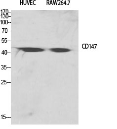
- Western Blot analysis of various cells using EMMPRIN Polyclonal Antibody diluted at 1:2000
.jpg)
- Western Blot analysis of HEPG2-UV cells using EMMPRIN Polyclonal Antibody diluted at 1:2000
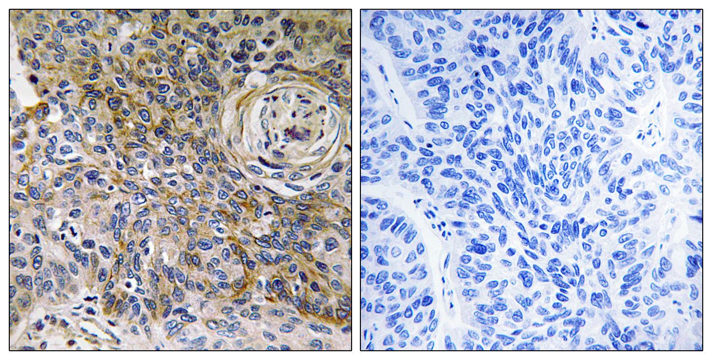
- Immunohistochemistry analysis of paraffin-embedded human breast carcinoma tissue, using CD147 Antibody. The picture on the right is blocked with the synthesized peptide.
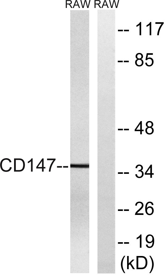
- Western blot analysis of lysates from RAW264.7 cells, using CD147 Antibody. The lane on the right is blocked with the synthesized peptide.


