EGFR Polyclonal Antibody
- Catalog No.:YT1497
- Applications:WB;IHC;IF;ELISA
- Reactivity:Human;Mouse;Rat
- Target:
- EGFR
- Fields:
- >>EGFR tyrosine kinase inhibitor resistance;>>Endocrine resistance;>>MAPK signaling pathway;>>ErbB signaling pathway;>>Ras signaling pathway;>>Rap1 signaling pathway;>>Calcium signaling pathway;>>HIF-1 signaling pathway;>>FoxO signaling pathway;>>Phospholipase D signaling pathway;>>Endocytosis;>>PI3K-Akt signaling pathway;>>Focal adhesion;>>Adherens junction;>>Gap junction;>>JAK-STAT signaling pathway;>>Regulation of actin cytoskeleton;>>GnRH signaling pathway;>>Estrogen signaling pathway;>>Oxytocin signaling pathway;>>Relaxin signaling pathway;>>Parathyroid hormone synthesis, secretion and action;>>Cushing syndrome;>>Epithelial cell signaling in Helicobacter pylori infection;>>Shigellosis;>>Hepatitis C;>>Human cytomegalovirus infection;>>Human papillomavirus infection;>>Coronavirus disease - COVID-19;>>Pathways in cancer;>>Proteoglycans in cancer;>>MicroRNAs in cancer;>>Chemical carcinogenesis - receptor activation;>>Chemical carcinogenesis - reactive oxygen species;>>Colorectal cance
- Gene Name:
- EGFR
- Protein Name:
- Epidermal growth factor receptor
- Human Gene Id:
- 1956
- Human Swiss Prot No:
- P00533
- Mouse Gene Id:
- 13649
- Mouse Swiss Prot No:
- Q01279
- Immunogen:
- The antiserum was produced against synthesized peptide derived from human EGFR. AA range:986-1035
- Specificity:
- EGFR Polyclonal Antibody detects endogenous levels of EGFR protein.
- Formulation:
- Liquid in PBS containing 50% glycerol, 0.5% BSA and 0.02% sodium azide.
- Source:
- Polyclonal, Rabbit,IgG
- Dilution:
- WB 1:500 - 1:2000. IHC 1:100 - 1:300. IF 1:200 - 1:1000. ELISA: 1:5000. Not yet tested in other applications.
- Purification:
- The antibody was affinity-purified from rabbit antiserum by affinity-chromatography using epitope-specific immunogen.
- Concentration:
- 1 mg/ml
- Storage Stability:
- -15°C to -25°C/1 year(Do not lower than -25°C)
- Other Name:
- EGFR;ERBB;ERBB1;HER1;Epidermal growth factor receptor;Proto-oncogene c-ErbB-1;Receptor tyrosine-protein kinase erbB-1
- Observed Band(KD):
- 175kD
- Background:
- The protein encoded by this gene is a transmembrane glycoprotein that is a member of the protein kinase superfamily. This protein is a receptor for members of the epidermal growth factor family. EGFR is a cell surface protein that binds to epidermal growth factor. Binding of the protein to a ligand induces receptor dimerization and tyrosine autophosphorylation and leads to cell proliferation. Mutations in this gene are associated with lung cancer. [provided by RefSeq, Jun 2016],
- Function:
- catalytic activity:ATP + a [protein]-L-tyrosine = ADP + a [protein]-L-tyrosine phosphate.,disease:Defects in EGFR are associated with lung cancer [MIM:211980].,function:Isoform 2/truncated isoform may act as an antagonist.,function:Receptor for EGF, but also for other members of the EGF family, as TGF-alpha, amphiregulin, betacellulin, heparin-binding EGF-like growth factor, GP30 and vaccinia virus growth factor. Is involved in the control of cell growth and differentiation. Phosphorylates MUC1 in breast cancer cells and increases the interaction of MUC1 with C-SRC and CTNNB1/beta-catenin.,miscellaneous:Binding of EGF to the receptor leads to dimerization, internalization of the EGF-receptor complex, induction of the tyrosine kinase activity, stimulation of cell DNA synthesis, and cell proliferation.,online information:EGFR entry,PTM:Monoubiquitinated and polyubiquitinated upon EGF stimu
- Subcellular Location:
- Cell membrane ; Single-pass type I membrane protein . Endoplasmic reticulum membrane ; Single-pass type I membrane protein. Golgi apparatus membrane; Single-pass type I membrane protein. Nucleus membrane; Single-pass type I membrane protein. Endosome . Endosome membrane. Nucleus . In response to EGF, translocated from the cell membrane to the nucleus via Golgi and ER (PubMed:20674546, PubMed:17909029). Endocytosed upon activation by ligand (PubMed:2790960, PubMed:17182860, PubMed:27153536, PubMed:17909029). Colocalized with GPER1 in the nucleus of estrogen agonist-induced cancer-associated fibroblasts (CAF) (PubMed:20551055). .; [Isoform 2]: Secreted.
- Expression:
- Ubiquitously expressed. Isoform 2 is also expressed in ovarian cancers.
Regulation of proliferation and cell cycle by protein regulator of cytokinesis 1 in oral squamous cell carcinoma. Cell Death & Disease 2018 May 11 WB Human 1:500 HSC-2 cell,Cal-27 cell
PTENα regulates endocytosis and modulates olfactory function. FASEB JOURNAL 2019 Jul 10 WB Human,Mouse 1:1000 olfactory bulb (OB) HEK293T cell
HSP90 inhibitor AUY922 can reverse Fulvestrant induced feedback reaction in human breast cancer cells. CANCER SCIENCE 2017 May 19 WB Human MCF-7 cell,T47D cell
Marine bromophenol bis(2,3-dibromo-4,5-dihydroxybenzyl) ether, represses angiogenesis in HUVEC cells and in zebrafish embryos via inhibiting the VEGF signal systems. BIOMEDICINE & PHARMACOTHERAPY 2015 Sep 07 WB Human HUVECs
Insulin-like growth factor II mRNA binding protein 3 regulates proliferation, invasion and migration of neuroendocrine cancer cells. International Journal of Clinical and Experimental Pathology Int J Clin Exp Patho. 2017; 10(10): 10269–10275 WB Mouse STC-1 cell
Combined caveolin‑1 and epidermal growth factor receptor expression as a prognostic marker for breast cancer. Oncology Letters Oncol Lett. 2018 Jun;15(6):9271-9282 IHC Human Breast cancer tissue
Marine bromophenol bis (2, 3-dibromo-4, 5-dihydroxybenzyl) ether, represses angiogenesis in HUVEC cells and in zebrafish embryos via inhibiting the VEGF signal systems." Biomedicine & Pharmacotherapy 75 (2015): 58-66.
Zhou, Rui, et al. "Epidermal growth factor (EGF) promotes human malignant glioma invasion by mediating secretion of human cytomegalovirus infected monocyte-derived macrophages." Precision Medicine 2 (2015).
Zhao, Junyu, et al. "MiR-133a-3p transferred by circulating microvesicles derived from myocardial ischemic preconditioning protects cardiomyocytes against hypoxia/reoxygenation injury." (2020).
PTENα regulates endocytosis and modulates olfactory function. FASEB JOURNAL 2019 Jul 10 WB Human,Mouse 1:1000 olfactory bulb (OB) HEK293T cell
GDF-15 Inhibits ADP-Induced Human Platelet Aggregation through the GFRAL/RET Signaling Complex Biomolecules Baikang Xie WB Human 1:1000 platelets,erythrocytes,leukocytes
- June 19-2018
- WESTERN IMMUNOBLOTTING PROTOCOL
- June 19-2018
- IMMUNOHISTOCHEMISTRY-PARAFFIN PROTOCOL
- June 19-2018
- IMMUNOFLUORESCENCE PROTOCOL
- September 08-2020
- FLOW-CYTOMEYRT-PROTOCOL
- May 20-2022
- Cell-Based ELISA│解您多样本WB检测之困扰
- July 13-2018
- CELL-BASED-ELISA-PROTOCOL-FOR-ACETYL-PROTEIN
- July 13-2018
- CELL-BASED-ELISA-PROTOCOL-FOR-PHOSPHO-PROTEIN
- July 13-2018
- Antibody-FAQs
- Products Images
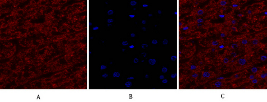
- Immunofluorescence analysis of human-liver tissue. 1,EGFR Polyclonal Antibody(red) was diluted at 1:200(4°C,overnight). 2, Cy3 labled Secondary antibody was diluted at 1:300(room temperature, 50min).3, Picture B: DAPI(blue) 10min. Picture A:Target. Picture B: DAPI. Picture C: merge of A+B

- Immunofluorescence analysis of mouse-liver tissue. 1,EGFR Polyclonal Antibody(red) was diluted at 1:200(4°C,overnight). 2, Cy3 labled Secondary antibody was diluted at 1:300(room temperature, 50min).3, Picture B: DAPI(blue) 10min. Picture A:Target. Picture B: DAPI. Picture C: merge of A+B

- Immunohistochemical analysis of paraffin-embedded Human-uterus tissue. 1,EGFR Polyclonal Antibody was diluted at 1:200(4°C,overnight). 2, Sodium citrate pH 6.0 was used for antibody retrieval(>98°C,20min). 3,Secondary antibody was diluted at 1:200(room tempeRature, 30min). Negative control was used by secondary antibody only.
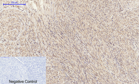
- Immunohistochemical analysis of paraffin-embedded Human-uterus-cancer tissue. 1,EGFR Polyclonal Antibody was diluted at 1:200(4°C,overnight). 2, Sodium citrate pH 6.0 was used for antibody retrieval(>98°C,20min). 3,Secondary antibody was diluted at 1:200(room tempeRature, 30min). Negative control was used by secondary antibody only.
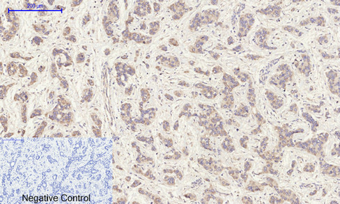
- Immunohistochemical analysis of paraffin-embedded Human-liver-cancer tissue. 1,EGFR Polyclonal Antibody was diluted at 1:200(4°C,overnight). 2, Sodium citrate pH 6.0 was used for antibody retrieval(>98°C,20min). 3,Secondary antibody was diluted at 1:200(room tempeRature, 30min). Negative control was used by secondary antibody only.

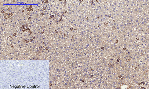
- Immunohistochemical analysis of paraffin-embedded Human-stomach tissue. 1,EGFR Polyclonal Antibody was diluted at 1:200(4°C,overnight). 2, Sodium citrate pH 6.0 was used for antibody retrieval(>98°C,20min). 3,Secondary antibody was diluted at 1:200(room tempeRature, 30min). Negative control was used by secondary antibody only.

- Immunohistochemical analysis of paraffin-embedded Human-stomach-cancer tissue. 1,EGFR Polyclonal Antibody was diluted at 1:200(4°C,overnight). 2, Sodium citrate pH 6.0 was used for antibody retrieval(>98°C,20min). 3,Secondary antibody was diluted at 1:200(room tempeRature, 30min). Negative control was used by secondary antibody only.
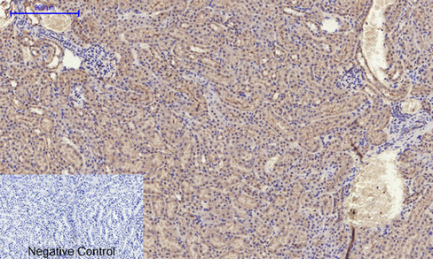
- Immunohistochemical analysis of paraffin-embedded Rat-liver tissue. 1,EGFR Polyclonal Antibody was diluted at 1:200(4°C,overnight). 2, Sodium citrate pH 6.0 was used for antibody retrieval(>98°C,20min). 3,Secondary antibody was diluted at 1:200(room tempeRature, 30min). Negative control was used by secondary antibody only.
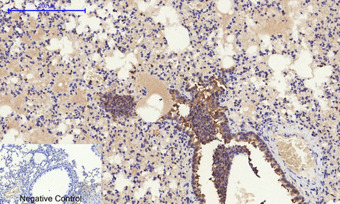
- Immunohistochemical analysis of paraffin-embedded Mouse-lung tissue. 1,EGFR Polyclonal Antibody was diluted at 1:200(4°C,overnight). 2, Sodium citrate pH 6.0 was used for antibody retrieval(>98°C,20min). 3,Secondary antibody was diluted at 1:200(room tempeRature, 30min). Negative control was used by secondary antibody only.
- Immunohistochemical analysis of paraffin-embedded Mouse-kidney tissue. 1,EGFR Polyclonal Antibody was diluted at 1:200(4°C,overnight). 2, Sodium citrate pH 6.0 was used for antibody retrieval(>98°C,20min). 3,Secondary antibody was diluted at 1:200(room tempeRature, 30min). Negative control was used by secondary antibody only.
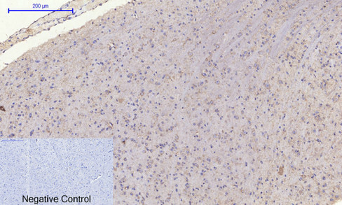
- Immunohistochemical analysis of paraffin-embedded Mouse-brain tissue. 1,EGFR Polyclonal Antibody was diluted at 1:200(4°C,overnight). 2, Sodium citrate pH 6.0 was used for antibody retrieval(>98°C,20min). 3,Secondary antibody was diluted at 1:200(room tempeRature, 30min). Negative control was used by secondary antibody only.

- Immunohistochemical analysis of paraffin-embedded Mouse-spleen tissue. 1,EGFR Polyclonal Antibody was diluted at 1:200(4°C,overnight). 2, Sodium citrate pH 6.0 was used for antibody retrieval(>98°C,20min). 3,Secondary antibody was diluted at 1:200(room tempeRature, 30min). Negative control was used by secondary antibody only.
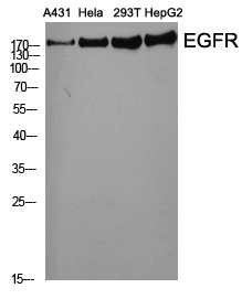
- Western Blot analysis of various cells using EGFR Polyclonal Antibody diluted at 1:2000
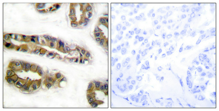
- Immunohistochemical analysis of paraffin-embedded Human breast cancer. Antibody was diluted at 1:100(4° overnight). High-pressure and temperature Tris-EDTA,pH8.0 was used for antigen retrieval. Negetive contrl (right) obtaned from antibody was pre-absorbed by immunogen peptide.
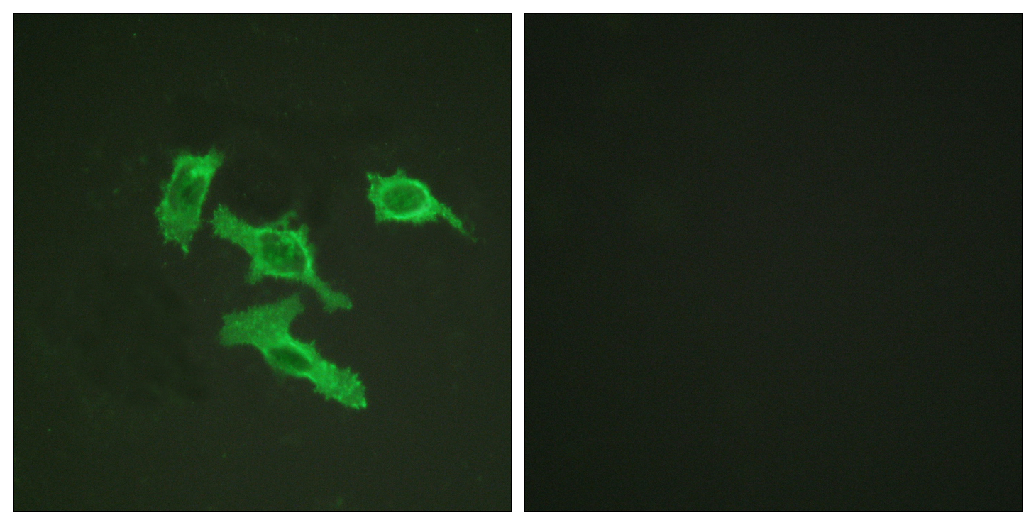
- Immunofluorescence analysis of HepG2 cells, using EGFR Antibody. The picture on the right is blocked with the synthesized peptide.


