EDG-8 Polyclonal Antibody
- Catalog No.:YT1472
- Applications:IHC;IF;ELISA
- Reactivity:Human;Rat;Mouse;
- Target:
- EDG-8
- Fields:
- >>Sphingolipid signaling pathway;>>Neuroactive ligand-receptor interaction
- Gene Name:
- S1PR5
- Protein Name:
- Sphingosine 1-phosphate receptor 5
- Human Gene Id:
- 53637
- Human Swiss Prot No:
- Q9H228
- Mouse Swiss Prot No:
- Q91X56
- Immunogen:
- The antiserum was produced against synthesized peptide derived from human EDG8. AA range:335-384
- Specificity:
- EDG-8 Polyclonal Antibody detects endogenous levels of EDG-8 protein.
- Formulation:
- Liquid in PBS containing 50% glycerol, 0.5% BSA and 0.02% sodium azide.
- Source:
- Polyclonal, Rabbit,IgG
- Dilution:
- IHC 1:100 - 1:300. IF 1:200 - 1:1000. ELISA: 1:5000. Not yet tested in other applications.
- Purification:
- The antibody was affinity-purified from rabbit antiserum by affinity-chromatography using epitope-specific immunogen.
- Concentration:
- 1 mg/ml
- Storage Stability:
- -15°C to -25°C/1 year(Do not lower than -25°C)
- Other Name:
- S1PR5;EDG8;Sphingosine 1-phosphate receptor 5;S1P receptor 5;S1P5;Endothelial differentiation G-protein-coupled receptor 8;Sphingosine 1-phosphate receptor Edg-8;S1P receptor Edg-8
- Molecular Weight(Da):
- 42kD
- Background:
- The lysosphingolipid sphingosine 1-phosphate (S1P) regulates cell proliferation, apoptosis, motility, and neurite retraction. Its actions may be both intracellular as a second messenger and extracellular as a receptor ligand. S1P and the structurally related lysolipid mediator lysophosphatidic acid (LPA) signal cells through a set of G protein-coupled receptors known as EDG receptors. Some EDG receptors (e.g., EDG1; MIM 601974) are S1P receptors; others (e.g., EDG2; MIM 602282) are LPA receptors.[supplied by OMIM, Mar 2008],
- Function:
- developmental stage:At 24 weeks of gestation, fragments of radial glial fibers are positive within the cortical plate and subplate of allocortical areas. These positive fragments often appear enlarged as varicosities and some of them terminate at blood vessels. Between 28 and 30 weeks of gestation, all iso- and allocortical areas contain immunolabelled radial glial fibers revealing curvature next to sulci. After 32 weeks of gestation, radial glial fibers gradually disappear; instead positive transitional stages between radial glia and astrocytes were found.,disease:Overexpressed in leukemic large granular lymphocyte (LGL). LGL is a lymphopropliferative disorder often associated with autoimmune disease.,function:Receptor for the lysosphingolipid sphingosine 1-phosphate (S1P). S1P is a bioactive lysophospholipid that elicits diverse physiological effect on most types of cells and tissues.
- Subcellular Location:
- Cell membrane; Multi-pass membrane protein.
- Expression:
- Widely expressed in the brain, most prominently in the corpus callosum, which is predominantly white matter. Detected in spleen, peripheral blood leukocytes, placenta, lung, aorta and fetal spleen. Low-level signal detected in many tissue extracts. Overexpressed in leukemic large granular lymphocytes. Isoform 1 is predominantly expressed in peripheral tissues. Isoform 2 is expressed in brain, spleen and peripheral blood leukocytes.
- June 19-2018
- WESTERN IMMUNOBLOTTING PROTOCOL
- June 19-2018
- IMMUNOHISTOCHEMISTRY-PARAFFIN PROTOCOL
- June 19-2018
- IMMUNOFLUORESCENCE PROTOCOL
- September 08-2020
- FLOW-CYTOMEYRT-PROTOCOL
- May 20-2022
- Cell-Based ELISA│解您多样本WB检测之困扰
- July 13-2018
- CELL-BASED-ELISA-PROTOCOL-FOR-ACETYL-PROTEIN
- July 13-2018
- CELL-BASED-ELISA-PROTOCOL-FOR-PHOSPHO-PROTEIN
- July 13-2018
- Antibody-FAQs
- Products Images
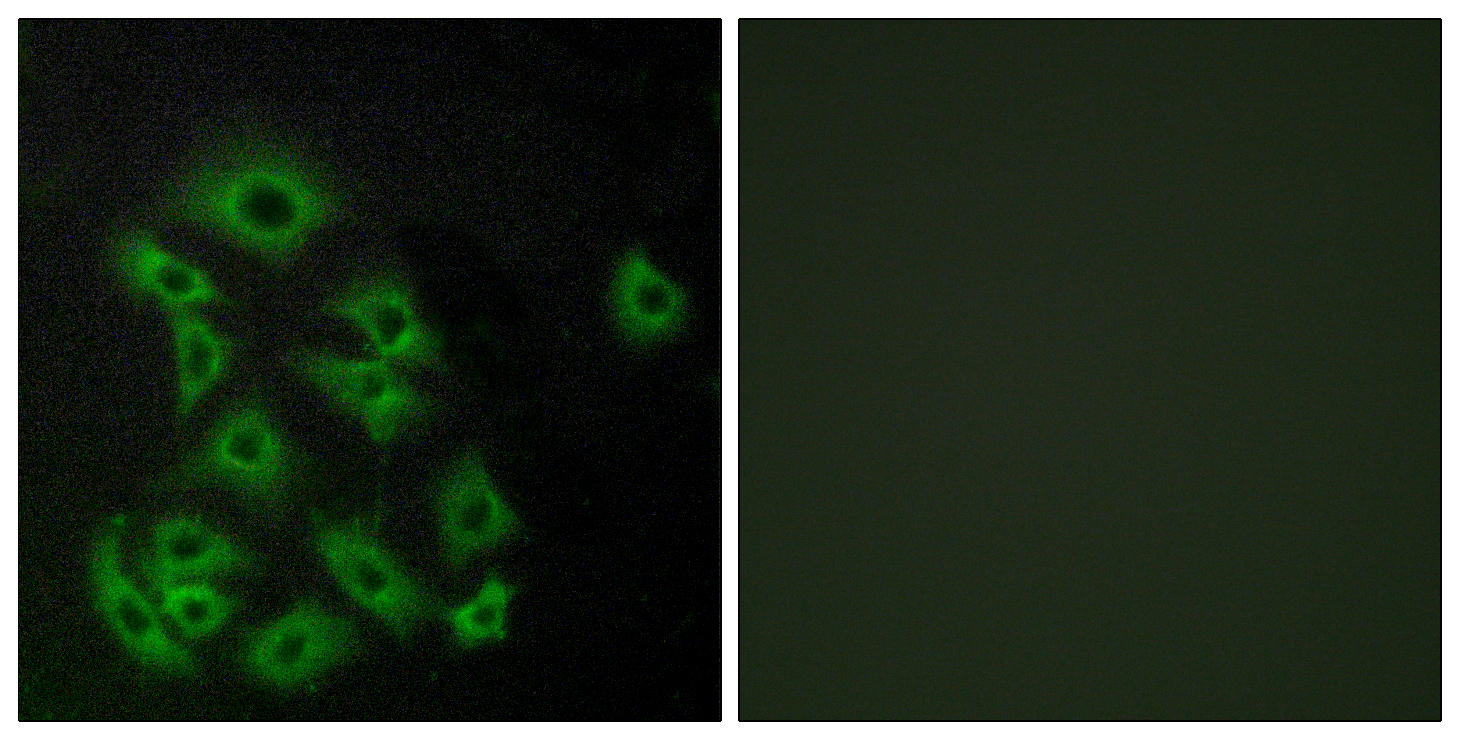
- Immunofluorescence analysis of A549 cells, using EDG8 Antibody. The picture on the right is blocked with the synthesized peptide.
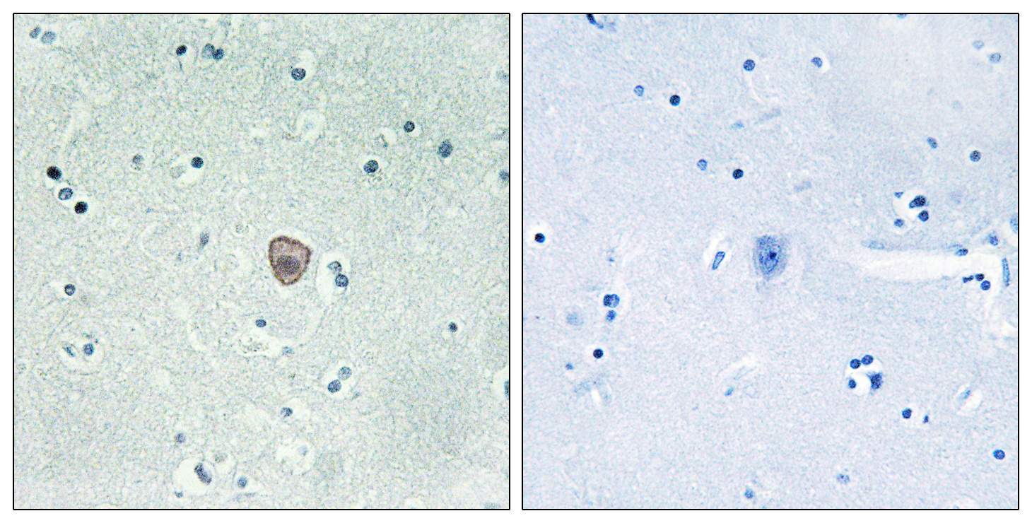
- Immunohistochemistry analysis of paraffin-embedded human brain tissue, using EDG8 Antibody. The picture on the right is blocked with the synthesized peptide.


