Dysferlin Polyclonal Antibody
- Catalog No.:YT1438
- Applications:WB;IF;ELISA
- Reactivity:Human;Mouse
- Target:
- Dysferlin
- Gene Name:
- DYSF
- Protein Name:
- Dysferlin
- Human Gene Id:
- 8291
- Human Swiss Prot No:
- O75923
- Mouse Gene Id:
- 26903
- Mouse Swiss Prot No:
- Q9ESD7
- Immunogen:
- The antiserum was produced against synthesized peptide derived from human Dysferlin. AA range:1981-2030
- Specificity:
- Dysferlin Polyclonal Antibody detects endogenous levels of Dysferlin protein.
- Formulation:
- Liquid in PBS containing 50% glycerol, 0.5% BSA and 0.02% sodium azide.
- Source:
- Polyclonal, Rabbit,IgG
- Dilution:
- WB 1:500 - 1:2000. IF 1:200 - 1:1000. ELISA: 1:10000. Not yet tested in other applications.
- Purification:
- The antibody was affinity-purified from rabbit antiserum by affinity-chromatography using epitope-specific immunogen.
- Concentration:
- 1 mg/ml
- Storage Stability:
- -15°C to -25°C/1 year(Do not lower than -25°C)
- Other Name:
- DYSF;FER1L1;Dysferlin;Dystrophy-associated fer-1-like protein;Fer-1-like protein 1
- Observed Band(KD):
- 240kD
- Background:
- dysferlin(DYSF) Homo sapiens The protein encoded by this gene belongs to the ferlin family and is a skeletal muscle protein found associated with the sarcolemma. It is involved in muscle contraction and contains C2 domains that play a role in calcium-mediated membrane fusion events, suggesting that it may be involved in membrane regeneration and repair. In addition, the protein encoded by this gene binds caveolin-3, a skeletal muscle membrane protein which is important in the formation of caveolae. Specific mutations in this gene have been shown to cause autosomal recessive limb girdle muscular dystrophy type 2B (LGMD2B) as well as Miyoshi myopathy. Alternative splicing results in multiple transcript variants. [provided by RefSeq, Aug 2008],
- Function:
- developmental stage:Expression in limb tissue from 5-6 weeks embryos; persists throughout development.,disease:Defects in DYSF are the cause of distal myopathy with anterior tibial onset (DMAT) [MIM:606768]. Onset of the disorder is between 14 and 28 years of age and the anterior tibial muscles are the first muscle group to be involved. Inheritance is autosomal recessive.,disease:Defects in DYSF are the cause of limb-girdle muscular dystrophy type 2B (LGMD2B) [MIM:253601]. LGMD2B is an autosomal recessive degenerative myopathy characterized by weakness and atrophy starting in the proximal pelvifemoral muscles, with onset in the late teens or later, massive elevation of serum creatine kinase levels and slow progression. Scapular muscle involvement is minor and not present at onset. Upper limb girdle involvement follows some years after the onset in lower limbs.,disease:Defects in DYSF are
- Subcellular Location:
- Cell membrane, sarcolemma; Single-pass type II membrane protein. Cytoplasmic vesicle membrane ; Single-pass type II membrane protein . Cell membrane. Colocalizes, during muscle differentiation, with BIN1 in the T-tubule system of myotubules and at the site of contact between two myotubes or a myoblast and a myotube. Wounding of myotubes led to its focal enrichment to the site of injury and to its relocalization in a Ca(2+)-dependent manner toward the plasma membrane. Colocalizes with AHNAK, AHNAK2 and PARVB at the sarcolemma of skeletal muscle. Detected on the apical plasma membrane of the syncytiotrophoblast. Reaches the plasmma membrane through a caveolin-independent mechanism. Retained by caveolin at the plasmma membrane (By similarity). Colocalizes, during muscle differentiation, with
- Expression:
- Expressed in skeletal muscle, myoblast, myotube and in the syncytiotrophoblast (STB) of the placenta (at protein level). Ubiquitous. Highly expressed in skeletal muscle. Also found in heart, brain, spleen, intestine, placenta and at lower levels in liver, lung, kidney and pancreas.
- June 19-2018
- WESTERN IMMUNOBLOTTING PROTOCOL
- June 19-2018
- IMMUNOHISTOCHEMISTRY-PARAFFIN PROTOCOL
- June 19-2018
- IMMUNOFLUORESCENCE PROTOCOL
- September 08-2020
- FLOW-CYTOMEYRT-PROTOCOL
- May 20-2022
- Cell-Based ELISA│解您多样本WB检测之困扰
- July 13-2018
- CELL-BASED-ELISA-PROTOCOL-FOR-ACETYL-PROTEIN
- July 13-2018
- CELL-BASED-ELISA-PROTOCOL-FOR-PHOSPHO-PROTEIN
- July 13-2018
- Antibody-FAQs
- Products Images
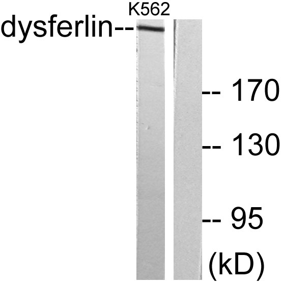
- Western blot analysis of lysates from K562 cells, using Dysferlin Antibody. The lane on the right is blocked with the synthesized peptide.


