Dyrk1A Polyclonal Antibody
- Catalog No.:YT1435
- Applications:WB;IHC;IF;ELISA
- Reactivity:Human;Mouse;Rat
- Target:
- DYR1A
- Gene Name:
- DYRK1A
- Protein Name:
- Dual specificity tyrosine-phosphorylation-regulated kinase 1A
- Human Gene Id:
- 1859
- Human Swiss Prot No:
- Q13627
- Mouse Gene Id:
- 13548
- Mouse Swiss Prot No:
- Q61214
- Rat Gene Id:
- 25255
- Rat Swiss Prot No:
- Q63470
- Immunogen:
- The antiserum was produced against synthesized peptide derived from human DYR1A. AA range:21-70
- Specificity:
- Dyrk1A Polyclonal Antibody detects endogenous levels of Dyrk1A protein.
- Formulation:
- Liquid in PBS containing 50% glycerol, 0.5% BSA and 0.02% sodium azide.
- Source:
- Polyclonal, Rabbit,IgG
- Dilution:
- WB 1:500 - 1:2000. IHC 1:100 - 1:300. IF 1:200 - 1:1000. ELISA: 1:20000. Not yet tested in other applications.
- Purification:
- The antibody was affinity-purified from rabbit antiserum by affinity-chromatography using epitope-specific immunogen.
- Concentration:
- 1 mg/ml
- Storage Stability:
- -15°C to -25°C/1 year(Do not lower than -25°C)
- Other Name:
- DYRK1A;DYRK;MNB;MNBH;Dual specificity tyrosine-phosphorylation-regulated kinase 1A;Dual specificity YAK1-related kinase;HP86;Protein kinase minibrain homolog;MNBH;hMNB
- Observed Band(KD):
- 90kD
- Background:
- This gene encodes a member of the Dual-specificity tyrosine phosphorylation-regulated kinase (DYRK) family. This member contains a nuclear targeting signal sequence, a protein kinase domain, a leucine zipper motif, and a highly conservative 13-consecutive-histidine repeat. It catalyzes its autophosphorylation on serine/threonine and tyrosine residues. It may play a significant role in a signaling pathway regulating cell proliferation and may be involved in brain development. This gene is a homolog of Drosophila mnb (minibrain) gene and rat Dyrk gene. It is localized in the Down syndrome critical region of chromosome 21, and is considered to be a strong candidate gene for learning defects associated with Down syndrome. Alternative splicing of this gene generates several transcript variants differing from each other either in the 5' UTR or in the 3' co
- Function:
- alternative products:Additional isoforms seem to exist,catalytic activity:ATP + a protein = ADP + a phosphoprotein.,developmental stage:Expressed in the developing central nervous system.,disease:Overexpressed 1.5-fold in fetal Down syndrome brain.,enzyme regulation:Inhibited by RANBP9.,function:May play a role in a signaling pathway regulating nuclear functions of cell proliferation. Phosphorylates serine, threonine and tyrosine residues in its sequence and in exogenous substrates.,PTM:Autophosphorylated on tyrosine residues.,similarity:Belongs to the protein kinase superfamily. CMGC Ser/Thr protein kinase family. MNB/DYRK subfamily.,similarity:Contains 1 protein kinase domain.,subunit:Interacts RAD54L2/ARIP4 (By similarity). Interacts with RANBP9.,tissue specificity:Ubiquitous. Highest levels in skeletal muscle, testis, fetal lung and fetal kidney.,
- Subcellular Location:
- Nucleus . Nucleus speckle .
- Expression:
- Ubiquitous. Highest levels in skeletal muscle, testis, fetal lung and fetal kidney.
MicroRNA-221-3p inhibits the inflammatory response of keratinocytes by regulating the DYRK1A/STAT3 signaling pathway to promote wound healing in diabetes Communications Biology Hu Keyan WB Human,Mouse skin wound tissue HaCaT cell
- June 19-2018
- WESTERN IMMUNOBLOTTING PROTOCOL
- June 19-2018
- IMMUNOHISTOCHEMISTRY-PARAFFIN PROTOCOL
- June 19-2018
- IMMUNOFLUORESCENCE PROTOCOL
- September 08-2020
- FLOW-CYTOMEYRT-PROTOCOL
- May 20-2022
- Cell-Based ELISA│解您多样本WB检测之困扰
- July 13-2018
- CELL-BASED-ELISA-PROTOCOL-FOR-ACETYL-PROTEIN
- July 13-2018
- CELL-BASED-ELISA-PROTOCOL-FOR-PHOSPHO-PROTEIN
- July 13-2018
- Antibody-FAQs
- Products Images

- Western Blot analysis of various cells using Dyrk1A Polyclonal Antibody diluted at 1:500 cells nucleus extracted by Minute TM Cytoplasmic and Nuclear Fractionation kit (SC-003,Inventbiotech,MN,USA).
.jpg)
- Western Blot analysis of HepG2 cells using Dyrk1A Polyclonal Antibody diluted at 1:500 cells nucleus extracted by Minute TM Cytoplasmic and Nuclear Fractionation kit (SC-003,Inventbiotech,MN,USA).
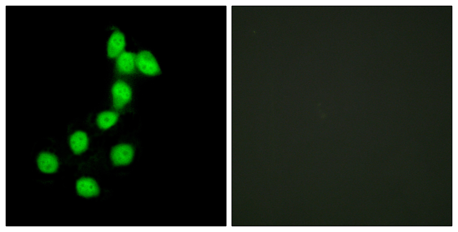
- Immunofluorescence analysis of HepG2 cells, using DYR1A Antibody. The picture on the right is blocked with the synthesized peptide.
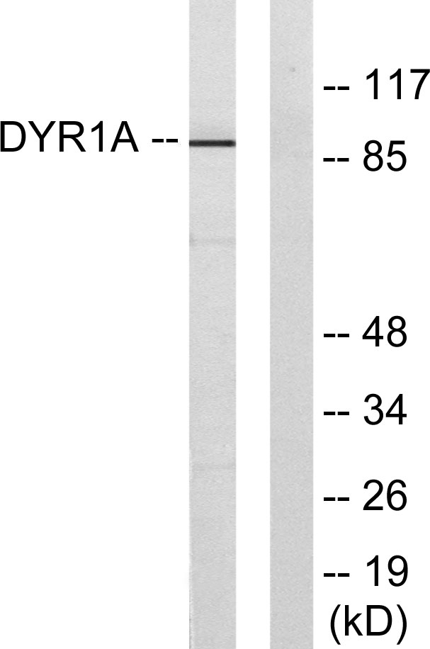
- Western blot analysis of lysates from HepG2 cells, using DYR1A Antibody. The lane on the right is blocked with the synthesized peptide.

- Immunohistochemical analysis of paraffin-embedded human Colon cancer. 1, Antibody was diluted at 1:200(4° overnight). 2, Tris-EDTA,pH9.0 was used for antigen retrieval. 3,Secondary antibody was diluted at 1:200(room temperature, 45min).


