DD3 Polyclonal Antibody
- Catalog No.:YT1306
- Applications:WB;ELISA
- Reactivity:Human
- Target:
- DD3
- Fields:
- >>Steroid hormone biosynthesis;>>Arachidonic acid metabolism;>>Folate biosynthesis;>>Metabolic pathways;>>Ovarian steroidogenesis;>>Chemical carcinogenesis - reactive oxygen species
- Gene Name:
- AKR1C3
- Protein Name:
- Aldo-keto reductase family 1 member C3
- Human Gene Id:
- 8644
- Human Swiss Prot No:
- P42330
- Immunogen:
- The antiserum was produced against synthesized peptide derived from human AKR1C3. AA range:191-240
- Specificity:
- DD3 Polyclonal Antibody detects endogenous levels of DD3 protein.
- Formulation:
- Liquid in PBS containing 50% glycerol, 0.5% BSA and 0.02% sodium azide.
- Source:
- Polyclonal, Rabbit,IgG
- Dilution:
- WB 1:500 - 1:2000. ELISA: 1:20000. Not yet tested in other applications.
- Purification:
- The antibody was affinity-purified from rabbit antiserum by affinity-chromatography using epitope-specific immunogen.
- Concentration:
- 1 mg/ml
- Storage Stability:
- -15°C to -25°C/1 year(Do not lower than -25°C)
- Other Name:
- AKR1C3;DDH1;HSD17B5;KIAA0119;PGFS;Aldo-keto reductase family 1 member C3;17-beta-hydroxysteroid dehydrogenase type 5;17-beta-HSD 5;3-alpha-HSD type II; brain;3-alpha-hydroxysteroid dehydrogenase type 2;3-alpha-HSD type 2;Chlordec
- Observed Band(KD):
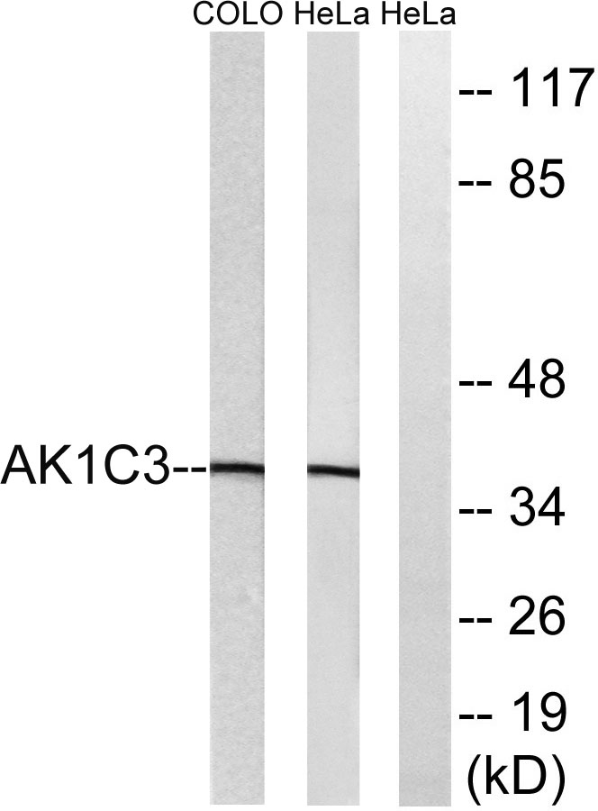
- 37kD
- Background:
- This gene encodes a member of the aldo/keto reductase superfamily, which consists of more than 40 known enzymes and proteins. These enzymes catalyze the conversion of aldehydes and ketones to their corresponding alcohols by utilizing NADH and/or NADPH as cofactors. The enzymes display overlapping but distinct substrate specificity. This enzyme catalyzes the reduction of prostaglandin (PG) D2, PGH2 and phenanthrenequinone (PQ), and the oxidation of 9alpha,11beta-PGF2 to PGD2. It may play an important role in the pathogenesis of allergic diseases such as asthma, and may also have a role in controlling cell growth and/or differentiation. This gene shares high sequence identity with three other gene members and is clustered with those three genes at chromosome 10p15-p14. Three transcript variants encoding different isoforms have been found for this gene. [provided by RefSeq, Dec 2011],
- Function:
- catalytic activity:(5Z,13E)-(15S)-9-alpha,11-alpha,15-trihydroxyprosta-5,13-dienoate + NADP(+) = (5Z,13E)-(15S)-9-alpha,15-dihydroxy-11-oxoprosta-5,13-dienoate + NADPH.,catalytic activity:Androsterone + NAD(P)(+) = 5-alpha-androstane-3,17-dione + NAD(P)H.,catalytic activity:Indan-1-ol + NAD(P)(+) = indanone + NAD(P)H.,catalytic activity:Testosterone + NAD(+) = androst-4-ene-3,17-dione + NADH.,catalytic activity:Testosterone + NADP(+) = androst-4-ene-3,17-dione + NADPH.,catalytic activity:Trans-1,2-dihydrobenzene-1,2-diol + NADP(+) = catechol + NADPH.,enzyme regulation:Strongly inhibited by nonsteroidal anti-inflammatory drugs (NSAID) including flufenamic acid and indomethacin. Also inhibited by the flavinoid, rutin, and by selective serotonin inhibitors (SSRIs).,function:Catalyzes the conversion of aldehydes and ketones to alcohols. Catalyzes the reduction of prostaglandin (PG) D2, PGH2
- Subcellular Location:
- Cytoplasm .
- Expression:
- Expressed in many tissues including adrenal gland, brain, kidney, liver, lung, mammary gland, placenta, small intestine, colon, spleen, prostate and testis. High expression in prostate and mammary gland. In the prostate, higher levels in epithelial cells than in stromal cells. In the brain, expressed in medulla, spinal cord, frontotemporal lobes, thalamus, subthalamic nuclei and amygdala. Weaker expression in the hippocampus, substantia nigra and caudate.
- June 19-2018
- WESTERN IMMUNOBLOTTING PROTOCOL
- June 19-2018
- IMMUNOHISTOCHEMISTRY-PARAFFIN PROTOCOL
- June 19-2018
- IMMUNOFLUORESCENCE PROTOCOL
- September 08-2020
- FLOW-CYTOMEYRT-PROTOCOL
- May 20-2022
- Cell-Based ELISA│解您多样本WB检测之困扰
- July 13-2018
- CELL-BASED-ELISA-PROTOCOL-FOR-ACETYL-PROTEIN
- July 13-2018
- CELL-BASED-ELISA-PROTOCOL-FOR-PHOSPHO-PROTEIN
- July 13-2018
- Antibody-FAQs
- Products Images

- Western blot analysis of lysates from HeLa and COLO cells, using AKR1C3 Antibody. The lane on the right is blocked with the synthesized peptide.


