Cyclin G Polyclonal Antibody
- Catalog No.:YT1181
- Applications:WB;IHC;IF;ELISA
- Reactivity:Human;Mouse;Rat
- Target:
- Cyclin G
- Fields:
- >>p53 signaling pathway;>>MicroRNAs in cancer
- Gene Name:
- CCNG1
- Protein Name:
- Cyclin-G1
- Human Gene Id:
- 900
- Human Swiss Prot No:
- P51959
- Mouse Gene Id:
- 12450
- Mouse Swiss Prot No:
- P51945
- Rat Gene Id:
- 25405
- Rat Swiss Prot No:
- P39950
- Immunogen:
- The antiserum was produced against synthesized peptide derived from human Cyclin G. AA range:161-210
- Specificity:
- Cyclin G Polyclonal Antibody detects endogenous levels of Cyclin G protein.
- Formulation:
- Liquid in PBS containing 50% glycerol, 0.5% BSA and 0.02% sodium azide.
- Source:
- Polyclonal, Rabbit,IgG
- Dilution:
- WB 1:500 - 1:2000. IHC 1:100 - 1:300. ELISA: 1:40000.. IF 1:50-200
- Purification:
- The antibody was affinity-purified from rabbit antiserum by affinity-chromatography using epitope-specific immunogen.
- Concentration:
- 1 mg/ml
- Storage Stability:
- -15°C to -25°C/1 year(Do not lower than -25°C)
- Other Name:
- CCNG1;CCNG;CYCG1;Cyclin-G1;Cyclin-G
- Observed Band(KD):
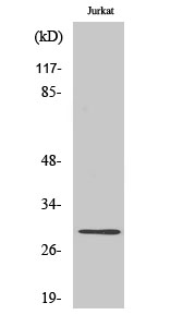
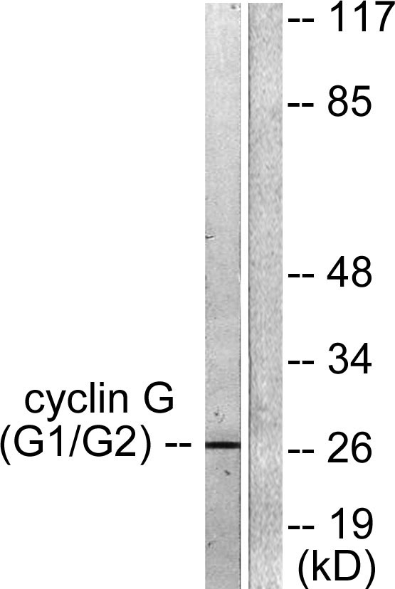
- 29kD
- Background:
- The eukaryotic cell cycle is governed by cyclin-dependent protein kinases (CDKs) whose activities are regulated by cyclins and CDK inhibitors. The protein encoded by this gene is a member of the cyclin family and contains the cyclin box. The encoded protein lacks the protein destabilizing (PEST) sequence that is present in other family members. Transcriptional activation of this gene can be induced by tumor protein p53. Two transcript variants encoding the same protein have been identified for this gene. [provided by RefSeq, Jul 2008],
- Function:
- developmental stage:Very low levels in normal cells during G1 phase, which increase as cells enter the S phase and stay high throughout the S and G2/M phases. In breast cancer cells consistent high levels are found throughout the cell cycle.,function:May play a role in growth regulation. Is associated with G2/M phase arrest in response to DNA damage. May be an intermediate by which p53 mediates its role as an inhibitor of cellular proliferation.,induction:Activated in breast and prostate cancer cells. Activated by actinomycin-D induced DNA damage.,similarity:Belongs to the cyclin family. Cyclin G subfamily.,subcellular location:DNA replication foci after DNA damage.,tissue specificity:High levels in skeletal muscle, ovary, kidney and colon.,
- Subcellular Location:
- Nucleus . DNA replication foci after DNA damage.
- Expression:
- High levels in skeletal muscle, ovary, kidney and colon.
- June 19-2018
- WESTERN IMMUNOBLOTTING PROTOCOL
- June 19-2018
- IMMUNOHISTOCHEMISTRY-PARAFFIN PROTOCOL
- June 19-2018
- IMMUNOFLUORESCENCE PROTOCOL
- September 08-2020
- FLOW-CYTOMEYRT-PROTOCOL
- May 20-2022
- Cell-Based ELISA│解您多样本WB检测之困扰
- July 13-2018
- CELL-BASED-ELISA-PROTOCOL-FOR-ACETYL-PROTEIN
- July 13-2018
- CELL-BASED-ELISA-PROTOCOL-FOR-PHOSPHO-PROTEIN
- July 13-2018
- Antibody-FAQs
- Products Images

- Western Blot analysis of various cells using Cyclin G Polyclonal Antibody diluted at 1:500 cells nucleus extracted by Minute TM Cytoplasmic and Nuclear Fractionation kit (SC-003,Inventbiotech,MN,USA).

- Immunohistochemistry analysis of paraffin-embedded human lung carcinoma tissue, using Cyclin G Antibody. The picture on the right is blocked with the synthesized peptide.

- Western blot analysis of lysates from Jurkat cells, using Cyclin G Antibody. The lane on the right is blocked with the synthesized peptide.


