Cyclin A Polyclonal Antibody
- Catalog No.:YT1167
- Applications:WB;IHC;IF;ELISA
- Reactivity:Human;Mouse;Rat;Monkey
- Target:
- Cyclin A
- Fields:
- >>Cell cycle;>>AMPK signaling pathway;>>Cellular senescence;>>Progesterone-mediated oocyte maturation;>>Hepatitis B;>>Human papillomavirus infection;>>Human T-cell leukemia virus 1 infection;>>Epstein-Barr virus infection;>>Pathways in cancer;>>Transcriptional misregulation in cancer;>>Viral carcinogenesis;>>Acute myeloid leukemia
- Gene Name:
- CCNA1/CCNA2
- Protein Name:
- Cyclin-A1/2
- Human Gene Id:
- 8900/890
- Human Swiss Prot No:
- P78396/P20248
- Mouse Gene Id:
- 12427/12428
- Rat Gene Id:
- 295052
- Rat Swiss Prot No:
- Q6AY13
- Immunogen:
- The antiserum was produced against synthesized peptide derived from human Cyclin A. AA range:221-270
- Specificity:
- Cyclin A Polyclonal Antibody detects endogenous levels of Cyclin A protein.
- Formulation:
- Liquid in PBS containing 50% glycerol, 0.5% BSA and 0.02% sodium azide.
- Source:
- Polyclonal, Rabbit,IgG
- Dilution:
- WB 1:500 - 1:2000. IHC 1:100 - 1:300. IF 1:200 - 1:1000. ELISA: 1:10000. Not yet tested in other applications.
- Purification:
- The antibody was affinity-purified from rabbit antiserum by affinity-chromatography using epitope-specific immunogen.
- Concentration:
- 1 mg/ml
- Storage Stability:
- -15°C to -25°C/1 year(Do not lower than -25°C)
- Other Name:
- CCNA1;Cyclin-A1;CCNA2;CCN1;CCNA;Cyclin-A2;Cyclin-A
- Observed Band(KD):
- 53kD
- Background:
- The protein encoded by this gene belongs to the highly conserved cyclin family, whose members are characterized by a dramatic periodicity in protein abundance through the cell cycle. Cyclins function as regulators of CDK kinases. Different cyclins exhibit distinct expression and degradation patterns which contribute to the temporal coordination of each mitotic event. The cyclin encoded by this gene was shown to be expressed in testis and brain, as well as in several leukemic cell lines, and is thought to primarily function in the control of the germline meiotic cell cycle. This cyclin binds both CDK2 and CDC2 kinases, which give two distinct kinase activities, one appearing in S phase, the other in G2, and thus regulate separate functions in cell cycle. This cyclin was found to bind to important cell cycle regulators, such as Rb family proteins, transcription factor E2F-1, and the p21 family proteins. Multi
- Function:
- developmental stage:Expression increases in early G1 phase and reaches highest levels during the S and G2/M phases.,function:May be involved in the control of the cell cycle at the G1/S (start) and G2/M (mitosis) transitions. May primarily function in the control of the germline meiotic cell cycle and additionally in the control of mitotic cell cycle in some somatic cells.,similarity:Belongs to the cyclin family.,similarity:Belongs to the cyclin family. Cyclin AB subfamily.,subunit:Interacts with the CDK2 and the CDC2 protein kinases to form a serine/threonine kinase holoenzyme complex. The cyclin subunit imparts substrate specificity to the complex. Does not bind CDK4 and CDK5 (in vitro). The cyclin A1-CDK2 complex interacts with transcription factor E2F-1 and RB proteins. Found in a complex with CDK2, CABLES1 and CCNE1 (By similarity). Interacts with INCA1 and KLHDC9.,tissue specificit
- Subcellular Location:
- Nucleus .
- Expression:
- Very high levels in testis and very low levels in brain. Also found in myeloid leukemia cell lines.
Melatonin treatment improves human umbilical cord mesenchymal stem cell therapy in a mouse model of type II diabetes mellitus via the PI3K/AKT signaling pathway. Stem Cell Research & Therapy2022 Dec;13(1):1-15. Human,Mouse 1:1200 liver tissue hUC-MSC
Flavonoid-Rich Extract of Oldenlandia diffusa (Willd.) Roxb. Inhibits Gastric Cancer by Activation of Caspase-Dependent Mitochondrial Apoptosis Chinese Journal of Integrative Medicine Qiu-lan Wang WB Human
- June 19-2018
- WESTERN IMMUNOBLOTTING PROTOCOL
- June 19-2018
- IMMUNOHISTOCHEMISTRY-PARAFFIN PROTOCOL
- June 19-2018
- IMMUNOFLUORESCENCE PROTOCOL
- September 08-2020
- FLOW-CYTOMEYRT-PROTOCOL
- May 20-2022
- Cell-Based ELISA│解您多样本WB检测之困扰
- July 13-2018
- CELL-BASED-ELISA-PROTOCOL-FOR-ACETYL-PROTEIN
- July 13-2018
- CELL-BASED-ELISA-PROTOCOL-FOR-PHOSPHO-PROTEIN
- July 13-2018
- Antibody-FAQs
- Products Images

- Immunofluorescence analysis of Hela cell. 1,Cyclin A Polyclonal Antibody(red) was diluted at 1:200(4° overnight). β-actin Monoclonal Antibody(5B7)(green) was diluted at 1:200(4° overnight). 2, Goat Anti Rabbit Alexa Fluor 594 Catalog:RS3611 was diluted at 1:1000(room temperature, 50min). Goat Anti Mouse Alexa Fluor 488 Catalog:RS3208 was diluted at 1:1000(room temperature, 50min).

- Immunofluorescence analysis of mouse-kidney tissue. 1,Cyclin A Polyclonal Antibody(red) was diluted at 1:200(4°C,overnight). 2, Cy3 labled Secondary antibody was diluted at 1:300(room temperature, 50min).3, Picture B: DAPI(blue) 10min. Picture A:Target. Picture B: DAPI. Picture C: merge of A+B
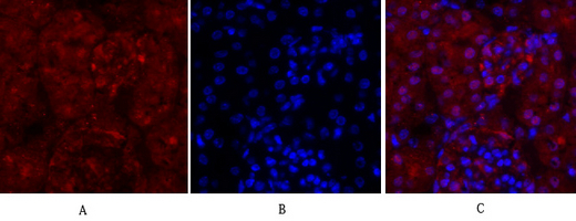
- Immunofluorescence analysis of mouse-kidney tissue. 1,Cyclin A Polyclonal Antibody(red) was diluted at 1:200(4°C,overnight). 2, Cy3 labled Secondary antibody was diluted at 1:300(room temperature, 50min).3, Picture B: DAPI(blue) 10min. Picture A:Target. Picture B: DAPI. Picture C: merge of A+B
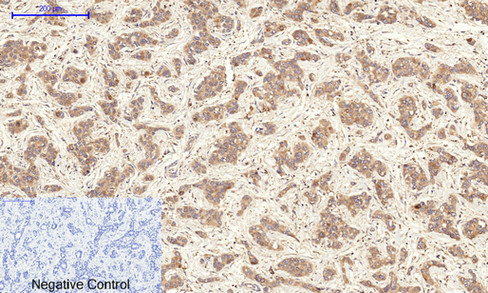
- Immunohistochemical analysis of paraffin-embedded Human-liver-cancer tissue. 1,Cyclin A Polyclonal Antibody was diluted at 1:200(4°C,overnight). 2, Sodium citrate pH 6.0 was used for antibody retrieval(>98°C,20min). 3,Secondary antibody was diluted at 1:200(room tempeRature, 30min). Negative control was used by secondary antibody only.
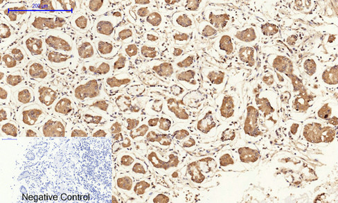
- Immunohistochemical analysis of paraffin-embedded Human-stomach tissue. 1,Cyclin A Polyclonal Antibody was diluted at 1:200(4°C,overnight). 2, Sodium citrate pH 6.0 was used for antibody retrieval(>98°C,20min). 3,Secondary antibody was diluted at 1:200(room tempeRature, 30min). Negative control was used by secondary antibody only.
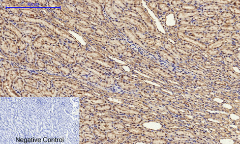
- Immunohistochemical analysis of paraffin-embedded Rat-kidney tissue. 1,Cyclin A Polyclonal Antibody was diluted at 1:200(4°C,overnight). 2, Sodium citrate pH 6.0 was used for antibody retrieval(>98°C,20min). 3,Secondary antibody was diluted at 1:200(room tempeRature, 30min). Negative control was used by secondary antibody only.
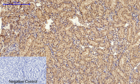
- Immunohistochemical analysis of paraffin-embedded Mouse-kidney tissue. 1,Cyclin A Polyclonal Antibody was diluted at 1:200(4°C,overnight). 2, Sodium citrate pH 6.0 was used for antibody retrieval(>98°C,20min). 3,Secondary antibody was diluted at 1:200(room tempeRature, 30min). Negative control was used by secondary antibody only.
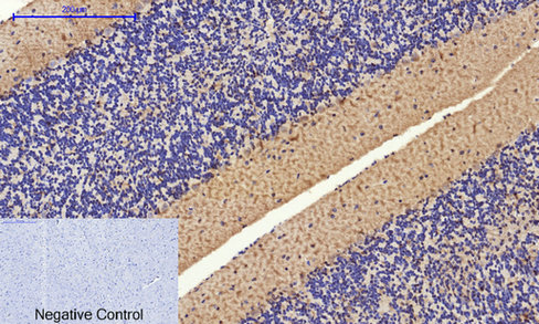
- Immunohistochemical analysis of paraffin-embedded Mouse-brain tissue. 1,Cyclin A Polyclonal Antibody was diluted at 1:200(4°C,overnight). 2, Sodium citrate pH 6.0 was used for antibody retrieval(>98°C,20min). 3,Secondary antibody was diluted at 1:200(room tempeRature, 30min). Negative control was used by secondary antibody only.
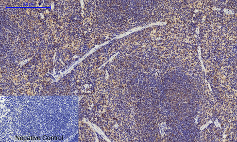
- Immunohistochemical analysis of paraffin-embedded Mouse-spleen tissue. 1,Cyclin A Polyclonal Antibody was diluted at 1:200(4°C,overnight). 2, Sodium citrate pH 6.0 was used for antibody retrieval(>98°C,20min). 3,Secondary antibody was diluted at 1:200(room tempeRature, 30min). Negative control was used by secondary antibody only.

- Immunohistochemical analysis of paraffin-embedded Rat-spinal-cord tissue. 1,Cyclin A Polyclonal Antibody was diluted at 1:200(4°C,overnight). 2, Sodium citrate pH 6.0 was used for antibody retrieval(>98°C,20min). 3,Secondary antibody was diluted at 1:200(room tempeRature, 30min). Negative control was used by secondary antibody only.
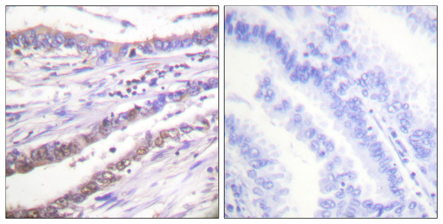
- Immunohistochemistry analysis of paraffin-embedded human lung carcinoma tissue, using Cyclin A Antibody. The picture on the right is blocked with the synthesized peptide.
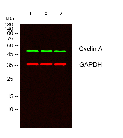
- Western blot analysis of lysates from 1) 3T3, 2) Jurkat, 3) K562 cells, (Green) primary antibody was diluted at 1:1000, 4°over night, secondary antibody(cat:RS23920)was diluted at 1:10000, 37° 1hour. (Red) GAPDH Monoclonal Antibody(2B8) (cat:YM3029) antibody was diluted at 1:5000 as loading control, 4° over night,secondary antibody(cat:RS23710)was diluted at 1:10000, 37° 1hour.


