COP1 Polyclonal Antibody
- Catalog No.:YT1058
- Applications:WB;ELISA
- Reactivity:Human;Mouse
- Target:
- COP1
- Fields:
- >>p53 signaling pathway;>>Ubiquitin mediated proteolysis
- Gene Name:
- RFWD2
- Protein Name:
- E3 ubiquitin-protein ligase RFWD2
- Human Gene Id:
- 64326
- Human Swiss Prot No:
- Q8NHY2
- Mouse Gene Id:
- 26374
- Mouse Swiss Prot No:
- Q9R1A8
- Immunogen:
- The antiserum was produced against synthesized peptide derived from human RFWD2. AA range:353-402
- Specificity:
- COP1 Polyclonal Antibody detects endogenous levels of COP1 protein.
- Formulation:
- Liquid in PBS containing 50% glycerol, 0.5% BSA and 0.02% sodium azide.
- Source:
- Polyclonal, Rabbit,IgG
- Dilution:
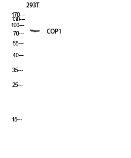
- WB 1:500 - 1:2000. ELISA: 1:40000. Not yet tested in other applications.
- Purification:
- The antibody was affinity-purified from rabbit antiserum by affinity-chromatography using epitope-specific immunogen.
- Concentration:
- 1 mg/ml
- Storage Stability:
- -15°C to -25°C/1 year(Do not lower than -25°C)
- Other Name:
- RFWD2;COP1;RNF200;E3 ubiquitin-protein ligase RFWD2;Constitutive photomorphogenesis protein 1 homolog;hCOP1;RING finger and WD repeat domain protein 2;RING finger protein 200
- Observed Band(KD):
- 110kD
- Background:
- domain:The RING finger domain, in addition to its role in ubiquitination, functions as a structural scaffold to bring two clusters of positive-charged residues within spatial proximity to mimic a bipartite nuclear localization signal (NLS).,function:E3 ubiquitin-protein ligase that mediates ubiquitination and subsequent proteasomal degradation of target proteins. E3 ubiquitin ligases accept ubiquitin from an E2 ubiquitin-conjugating enzyme in the form of a thioester and then directly transfers the ubiquitin to targeted substrates. Involved in JUN ubiquitination and degradation. Directly involved in p53 (TP53) ubiquitination and degradation, thereby abolishing p53-dependent transcription and apoptosis. Ubiquitinates p53 independently of MDM2 or RCHY1. Probably mediates E3 ubiquitin ligase activity by functioning as the essential RING domain subunit of larger E3 complexes. In contrast, it does not constitute the catalytic RING subunit in the DCX DET1-COP1 complex that negatively regulates JUN, the ubiquitin ligase activity being mediated by RBX1.,induction:By p53/TP53.,pathway:Protein modification; protein ubiquitination.,similarity:Belongs to the COP1 family.,similarity:Contains 1 RING-type zinc finger.,similarity:Contains 7 WD repeats.,subcellular location:In the nucleus, it forms nuclear speckles.,subunit:Homodimer. Homodimerization is mediated by the coiled coil domain. Component of the DCX DET1-COP1 ubiquitin ligase complex at least composed of RBX1, DET1, DDB1, CUL4A and COP1. Isoform 2 does not interact with CUL4A but still binds to RBX1, suggesting that the interaction may be mediated by another culllin protein. Isoform 1 and isoform 2 interact with CUL5 but not with CUL1, CUL2 not CUL3. Interacts with bZIP transcription factors JUN, JUNB and JUND but not with FOS, ATF2 nor XBP1. Interacts with p53 (TP53).,tissue specificity:Ubiquitously expressed at low level. Expressed at higher level in testis, placenta, skeletal muscle and heart.,
- Function:
- domain:The RING finger domain, in addition to its role in ubiquitination, functions as a structural scaffold to bring two clusters of positive-charged residues within spatial proximity to mimic a bipartite nuclear localization signal (NLS).,function:E3 ubiquitin-protein ligase that mediates ubiquitination and subsequent proteasomal degradation of target proteins. E3 ubiquitin ligases accept ubiquitin from an E2 ubiquitin-conjugating enzyme in the form of a thioester and then directly transfers the ubiquitin to targeted substrates. Involved in JUN ubiquitination and degradation. Directly involved in p53 (TP53) ubiquitination and degradation, thereby abolishing p53-dependent transcription and apoptosis. Ubiquitinates p53 independently of MDM2 or RCHY1. Probably mediates E3 ubiquitin ligase activity by functioning as the essential RING domain subunit of larger E3 complexes. In contrast, it
- Subcellular Location:
- Nucleus speckle. Cytoplasm. In the nucleus, it forms nuclear speckles.
- Expression:
- Ubiquitously expressed at low level. Expressed at higher level in testis, placenta, skeletal muscle and heart.
- June 19-2018
- WESTERN IMMUNOBLOTTING PROTOCOL
- June 19-2018
- IMMUNOHISTOCHEMISTRY-PARAFFIN PROTOCOL
- June 19-2018
- IMMUNOFLUORESCENCE PROTOCOL
- September 08-2020
- FLOW-CYTOMEYRT-PROTOCOL
- May 20-2022
- Cell-Based ELISA│解您多样本WB检测之困扰
- July 13-2018
- CELL-BASED-ELISA-PROTOCOL-FOR-ACETYL-PROTEIN
- July 13-2018
- CELL-BASED-ELISA-PROTOCOL-FOR-PHOSPHO-PROTEIN
- July 13-2018
- Antibody-FAQs
- Products Images

- Western blot analysis of 293T lysis using COP1 antibody. Antibody was diluted at 1:1000


