COL2A1 Polyclonal Antibody
- Catalog No.:YT1022
- Applications:WB;IHC;IF;ELISA
- Reactivity:Human;Mouse;Rat
- Target:
- Collagen II
- Fields:
- >>PI3K-Akt signaling pathway;>>Focal adhesion;>>ECM-receptor interaction;>>Protein digestion and absorption;>>Human papillomavirus infection
- Gene Name:
- COL2A1
- Protein Name:
- Collagen alpha-1(II) chain
- Human Gene Id:
- 1280
- Human Swiss Prot No:
- P02458
- Mouse Gene Id:
- 12824
- Mouse Swiss Prot No:
- P28481
- Rat Gene Id:
- 25412
- Rat Swiss Prot No:
- P05539
- Immunogen:
- The antiserum was produced against synthesized peptide derived from human Collagen II. AA range:101-150
- Specificity:
- COL2A1 Polyclonal Antibody detects endogenous levels of COL2A1 protein.
- Formulation:
- Liquid in PBS containing 50% glycerol, 0.5% BSA and 0.02% sodium azide.
- Source:
- Polyclonal, Rabbit,IgG
- Dilution:
- WB 1:500 - 1:2000. IHC 1:100 - 1:300. IF 1:200 - 1:1000. ELISA: 1:20000. Not yet tested in other applications.
- Purification:
- The antibody was affinity-purified from rabbit antiserum by affinity-chromatography using epitope-specific immunogen.
- Concentration:
- 1 mg/ml
- Storage Stability:
- -15°C to -25°C/1 year(Do not lower than -25°C)
- Other Name:
- COL2A1;Collagen alpha-1(II) chain;Alpha-1 type II collagen
- Observed Band(KD):
- 140kD
- Background:
- This gene encodes the alpha-1 chain of type II collagen, a fibrillar collagen found in cartilage and the vitreous humor of the eye. Mutations in this gene are associated with achondrogenesis, chondrodysplasia, early onset familial osteoarthritis, SED congenita, Langer-Saldino achondrogenesis, Kniest dysplasia, Stickler syndrome type I, and spondyloepimetaphyseal dysplasia Strudwick type. In addition, defects in processing chondrocalcin, a calcium binding protein that is the C-propeptide of this collagen molecule, are also associated with chondrodysplasia. There are two transcripts identified for this gene. [provided by RefSeq, Jul 2008],
- Function:
- disease:Defects in COL2A1 are a cause of primary avascular necrosis of femoral head (ANFH) [MIM:608805]; also called ischemic necrosis of the femoral head or osteonecrosis of the femoral head. ANFH causes disability that often requires surgical intervention. Most cases are sporadic, but families in which there is an autosomal dominant inheritance of the disease have been identified. It has been estimated that 300,000 to 600,000 people in the United States have ANFH. Approximately 15,000 new cases of this common and disabling disorder are reported annually. The age at the onset is earlier than that for osteoarthritis. The diagnosis is typically made when patients are between the ages of 30 and 60 years. The clinical manifestations, such as pain on exertion, a limping gait, and a discrepancy in leg length, cause considerable disability. Moreover, nearly 10 percent of the 500,000 total-hip
- Subcellular Location:
- Secreted, extracellular space, extracellular matrix .
- Expression:
- Isoform 2 is highly expressed in juvenile chondrocyte and low in fetal chondrocyte.
Bromodomain-containing protein 7 regulates matrix metabolism and apoptosis in human nucleus pulposus cells through the BRD7-PI3K-YAP1 signaling axis. EXPERIMENTAL CELL RESEARCH Exp Cell Res. 2021 Aug;405:112658 IF Human 1 : 100 Nucleus pulposus (NP) cell
The role of SIRT1 in BMP2-induced chondrogenic differentiation and cartilage maintenance under oxidative stress.. Aging-US Aging-Us. 2020 May 31; 12(10): 9000–9013 WB Mouse 1:500 C3H10T1/2 cells
Atorvastatin inhibited TNF-α induced matrix degradation in rat nucleus pulposus cells by suppressing NLRP3 inflammasome activity and inducing autophagy through NF-κB signaling. CELL CYCLE Cell Cycle. 2021;20(20):2160-2173 IF Rat 1:100 NP cell
Long non-coding RNA MEG3 inhibits chondrogenic differentiation of synovium-derived mesenchymal stem cells by epigenetically inhibiting TRIB2 via methyltransferase EZH2. CELLULAR SIGNALLING Cell Signal. 2019 Nov;63:109379 WB,IF Human synovium-derived mesenchymal stem cells(SMSCs)
Lycopene alleviates disc degeneration under oxidative stress through the Nrf2 signaling pathway. MOLECULAR AND CELLULAR PROBES Mol Cell Probe. 2020 Jun;51:101559 WB Human 1:500 NPCs
Koumine inhibits IL-1β-induced chondrocyte inflammation and ameliorates extracellular matrix degradation in osteoarthritic cartilage through activation of PINK1/Parkin-mediated mitochondrial autophagy BIOMEDICINE & PHARMACOTHERAPY Xiangyi Kong IHC,WB Rat 1:200 articular cartilage tissue RCCS-1 cell
Diagnostic value of serum COMP and ADAMTS7 for intervertebral disc degeneration EUROPEAN JOURNAL OF MEDICAL RESEARCH Ding Jing-Yu WB Human 1:1000 nucleus pulposus (NP) cell
Unlocking the potential of β-1,3-xylooligosaccharides from Caulerpa lentillifera: structural characterization, antioxidative and anti-osteoarthritis applications Chemical and Biological Technologies in Agriculture Cai Lixi WB Rat 1:1000 chondrocyte
- June 19-2018
- WESTERN IMMUNOBLOTTING PROTOCOL
- June 19-2018
- IMMUNOHISTOCHEMISTRY-PARAFFIN PROTOCOL
- June 19-2018
- IMMUNOFLUORESCENCE PROTOCOL
- September 08-2020
- FLOW-CYTOMEYRT-PROTOCOL
- May 20-2022
- Cell-Based ELISA│解您多样本WB检测之困扰
- July 13-2018
- CELL-BASED-ELISA-PROTOCOL-FOR-ACETYL-PROTEIN
- July 13-2018
- CELL-BASED-ELISA-PROTOCOL-FOR-PHOSPHO-PROTEIN
- July 13-2018
- Antibody-FAQs
- Products Images
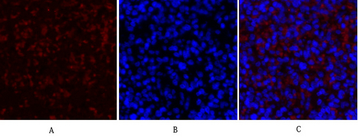
- Immunofluorescence analysis of rat-spleen tissue. 1,COL2A1 Polyclonal Antibody(red) was diluted at 1:200(4°C,overnight). 2, Cy3 labled Secondary antibody was diluted at 1:300(room temperature, 50min).3, Picture B: DAPI(blue) 10min. Picture A:Target. Picture B: DAPI. Picture C: merge of A+B
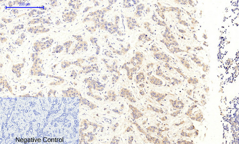
- Immunohistochemical analysis of paraffin-embedded Human-liver-cancer tissue. 1,COL2A1 Polyclonal Antibody was diluted at 1:200(4°C,overnight). 2, Sodium citrate pH 6.0 was used for antibody retrieval(>98°C,20min). 3,Secondary antibody was diluted at 1:200(room tempeRature, 30min). Negative control was used by secondary antibody only.
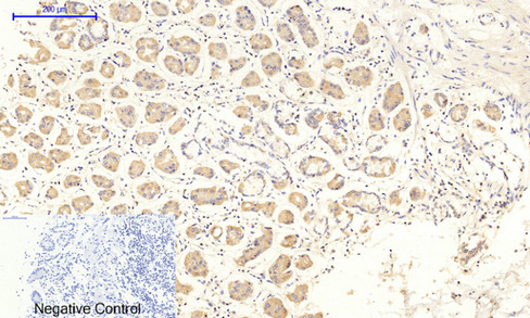
- Immunohistochemical analysis of paraffin-embedded Human-stomach tissue. 1,COL2A1 Polyclonal Antibody was diluted at 1:200(4°C,overnight). 2, Sodium citrate pH 6.0 was used for antibody retrieval(>98°C,20min). 3,Secondary antibody was diluted at 1:200(room tempeRature, 30min). Negative control was used by secondary antibody only.
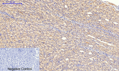
- Immunohistochemical analysis of paraffin-embedded Rat-kidney tissue. 1,COL2A1 Polyclonal Antibody was diluted at 1:200(4°C,overnight). 2, Sodium citrate pH 6.0 was used for antibody retrieval(>98°C,20min). 3,Secondary antibody was diluted at 1:200(room tempeRature, 30min). Negative control was used by secondary antibody only.
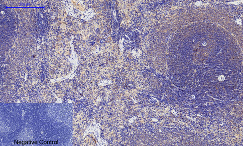
- Immunohistochemical analysis of paraffin-embedded Rat-spleen tissue. 1,COL2A1 Polyclonal Antibody was diluted at 1:200(4°C,overnight). 2, Sodium citrate pH 6.0 was used for antibody retrieval(>98°C,20min). 3,Secondary antibody was diluted at 1:200(room tempeRature, 30min). Negative control was used by secondary antibody only.
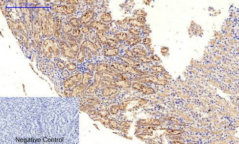
- Immunohistochemical analysis of paraffin-embedded Mouse-kidney tissue. 1,COL2A1 Polyclonal Antibody was diluted at 1:200(4°C,overnight). 2, Sodium citrate pH 6.0 was used for antibody retrieval(>98°C,20min). 3,Secondary antibody was diluted at 1:200(room tempeRature, 30min). Negative control was used by secondary antibody only.
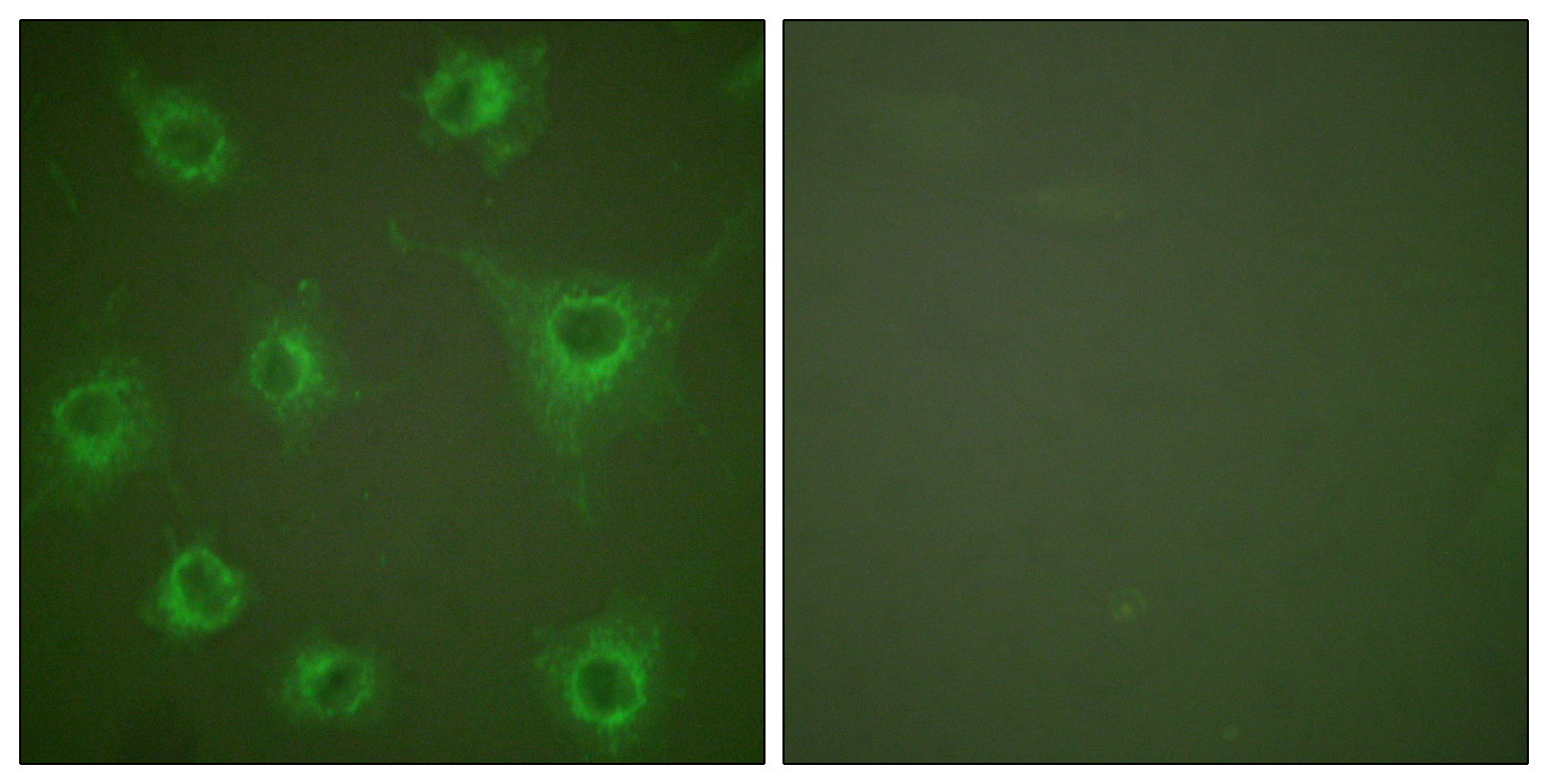
- Immunofluorescence analysis of COS7 cells, using Collagen II Antibody. The picture on the right is blocked with the synthesized peptide.
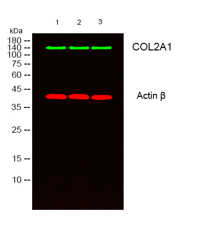
- Western blot analysis of lysates from 1) k562 , 2) COLO205,3) MCF-7 cells, (Green) primary antibody was diluted at 1:1000, 4°over night, secondary antibody(cat:RS23920)was diluted at 1:10000, 37° 1hour. (Red) Actin β Monoclonal Antibody(5B7) (cat:YM3028) antibody was diluted at 1:5000 as loading control, 4° over night,secondary antibody(cat:RS23710)was diluted at 1:10000, 37° 1hour.


