Cofilin Polyclonal Antibody
- Catalog No.:YT1007
- Applications:WB;IF;ELISA
- Reactivity:Human;Mouse;Rat
- Target:
- Cofilin
- Fields:
- >>Axon guidance;>>Fc gamma R-mediated phagocytosis;>>Regulation of actin cytoskeleton;>>Pertussis;>>Human immunodeficiency virus 1 infection
- Gene Name:
- CFL1/CFL2
- Protein Name:
- Cofilin-1/2
- Human Gene Id:
- 1073/1072
- Human Swiss Prot No:
- P23528/Q9Y281
- Mouse Gene Id:
- 12631/12632
- Rat Gene Id:
- 29271
- Rat Swiss Prot No:
- P45592
- Immunogen:
- The antiserum was produced against synthesized peptide derived from human Cofilin. AA range:61-110
- Specificity:
- Cofilin Polyclonal Antibody detects endogenous levels of Cofilin protein.
- Formulation:
- Liquid in PBS containing 50% glycerol, 0.5% BSA and 0.02% sodium azide.
- Source:
- Polyclonal, Rabbit,IgG
- Dilution:
- WB 1:500 - 1:2000. IF 1:200 - 1:1000. ELISA: 1:40000. Not yet tested in other applications.
- Purification:
- The antibody was affinity-purified from rabbit antiserum by affinity-chromatography using epitope-specific immunogen.
- Concentration:
- 1 mg/ml
- Storage Stability:
- -15°C to -25°C/1 year(Do not lower than -25°C)
- Other Name:
- CFL1;CFL;Cofilin-1;18 kDa phosphoprotein;p18;Cofilin; non-muscle isoform;CFL2;Cofilin-2;Cofilin, muscle isoform
- Observed Band(KD):
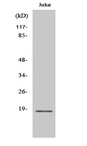
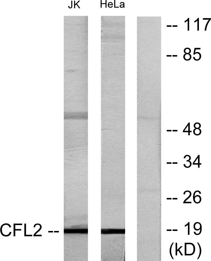
- 19kD
- Background:
- cofilin 1(CFL1) Homo sapiens The protein encoded by this gene can polymerize and depolymerize F-actin and G-actin in a pH-dependent manner. Increased phosphorylation of this protein by LIM kinase aids in Rho-induced reorganization of the actin cytoskeleton. Cofilin is a widely distributed intracellular actin-modulating protein that binds and depolymerizes filamentous F-actin and inhibits the polymerization of monomeric G-actin in a pH-dependent manner. It is involved in the translocation of actin-cofilin complex from cytoplasm to nucleus.[supplied by OMIM, Apr 2004],
- Function:
- function:Controls reversibly actin polymerization and depolymerization in a pH-sensitive manner. It has the ability to bind G- and F-actin in a 1:1 ratio of cofilin to actin. It is the major component of intranuclear and cytoplasmic actin rods.,online information:Cofilin entry,PTM:Phosphorylated on Ser-3 in resting cells.,similarity:Belongs to the actin-binding proteins ADF family.,similarity:Contains 1 ADF-H domain.,subcellular location:Almost completely in nucleus in cells exposed to heat shock or 10% dimethyl sulfoxide.,tissue specificity:Widely distributed in various tissues.,
- Subcellular Location:
- Nucleus matrix . Cytoplasm, cytoskeleton . Cell projection, ruffle membrane ; Peripheral membrane protein ; Cytoplasmic side . Cell projection, lamellipodium membrane ; Peripheral membrane protein ; Cytoplasmic side . Cell projection, lamellipodium . Cell projection, growth cone . Cell projection, axon . Colocalizes with the actin cytoskeleton in membrane ruffles and lamellipodia. Detected at the cleavage furrow and contractile ring during cytokinesis. Almost completely in nucleus in cells exposed to heat shock or 10% dimethyl sulfoxide.
- Expression:
- Widely distributed in various tissues.
- June 19-2018
- WESTERN IMMUNOBLOTTING PROTOCOL
- June 19-2018
- IMMUNOHISTOCHEMISTRY-PARAFFIN PROTOCOL
- June 19-2018
- IMMUNOFLUORESCENCE PROTOCOL
- September 08-2020
- FLOW-CYTOMEYRT-PROTOCOL
- May 20-2022
- Cell-Based ELISA│解您多样本WB检测之困扰
- July 13-2018
- CELL-BASED-ELISA-PROTOCOL-FOR-ACETYL-PROTEIN
- July 13-2018
- CELL-BASED-ELISA-PROTOCOL-FOR-PHOSPHO-PROTEIN
- July 13-2018
- Antibody-FAQs
- Products Images

- Western Blot analysis of various cells using Cofilin Polyclonal Antibody diluted at 1:1000

- Western blot analysis of lysates from Jurkat and HeLa cells, using Cofilin Antibody. The lane on the right is blocked with the synthesized peptide.


