CLCC1 Polyclonal Antibody
- Catalog No.:YT0962
- Applications:WB;IF;ELISA
- Reactivity:Human;Rat;Mouse;
- Target:
- CLCC1
- Gene Name:
- CLCC1
- Protein Name:
- Chloride channel CLIC-like protein 1
- Human Gene Id:
- 23155
- Human Swiss Prot No:
- Q96S66
- Mouse Swiss Prot No:
- Q99LI2
- Immunogen:
- The antiserum was produced against synthesized peptide derived from human CLCC1. AA range:391-440
- Specificity:
- CLCC1 Polyclonal Antibody detects endogenous levels of CLCC1 protein.
- Formulation:
- Liquid in PBS containing 50% glycerol, 0.5% BSA and 0.02% sodium azide.
- Source:
- Polyclonal, Rabbit,IgG
- Dilution:
- WB 1:500 - 1:2000. IF 1:200 - 1:1000. ELISA: 1:40000. Not yet tested in other applications.
- Purification:
- The antibody was affinity-purified from rabbit antiserum by affinity-chromatography using epitope-specific immunogen.
- Concentration:
- 1 mg/ml
- Storage Stability:
- -15°C to -25°C/1 year(Do not lower than -25°C)
- Other Name:
- CLCC1;KIAA0761;MCLC;Chloride channel CLIC-like protein 1;Mid-1-related chloride channel protein 1
- Observed Band(KD):
- 62kD
- Background:
- function:Seems to act as a chloride ion channel.,similarity:Belongs to the chloride channel MCLC family.,
- Function:
- function:Seems to act as a chloride ion channel.,similarity:Belongs to the chloride channel MCLC family.,
- Subcellular Location:
- Endoplasmic reticulum membrane ; Multi-pass membrane protein . Golgi apparatus membrane ; Multi-pass membrane protein . Nucleus membrane ; Multi-pass membrane protein . Within the endoplasmic reticulum (ER), localizes to the mitochondria-associated ER membrane, a zone of contact between the ER and mitochondrial membranes. .
- Expression:
- Expressed in the retina of the eye, with extensive expression in the lamina cribrosa, optic nerve, ganglion cell layer, inner nuclear layer, outer nuclear layer and retinal pigment epithelium.
- June 19-2018
- WESTERN IMMUNOBLOTTING PROTOCOL
- June 19-2018
- IMMUNOHISTOCHEMISTRY-PARAFFIN PROTOCOL
- June 19-2018
- IMMUNOFLUORESCENCE PROTOCOL
- September 08-2020
- FLOW-CYTOMEYRT-PROTOCOL
- May 20-2022
- Cell-Based ELISA│解您多样本WB检测之困扰
- July 13-2018
- CELL-BASED-ELISA-PROTOCOL-FOR-ACETYL-PROTEIN
- July 13-2018
- CELL-BASED-ELISA-PROTOCOL-FOR-PHOSPHO-PROTEIN
- July 13-2018
- Antibody-FAQs
- Products Images
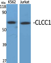
- Western Blot analysis of various cells using CLCC1 Polyclonal Antibody diluted at 1:1000
.jpg)
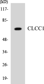
- Western Blot analysis of HepG2 cells using CLCC1 Polyclonal Antibody diluted at 1:1000
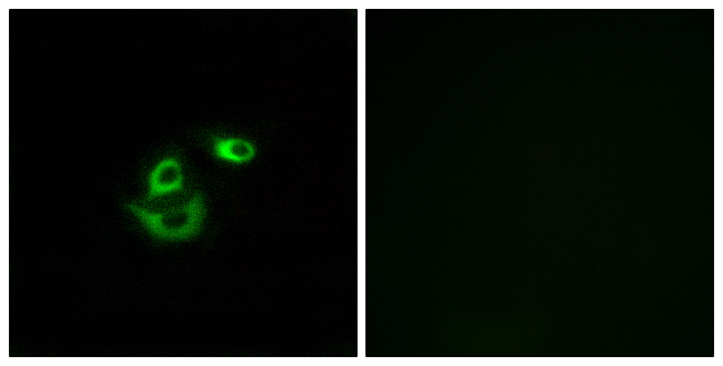
- Immunofluorescence analysis of A549 cells, using CLCC1 Antibody. The picture on the right is blocked with the synthesized peptide.
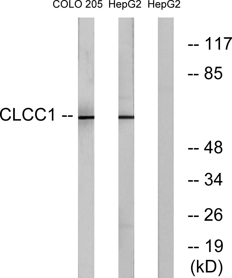
- Western blot analysis of lysates from COLO and HepG2 cells, using CLCC1 Antibody. The lane on the right is blocked with the synthesized peptide.
- Western blot analysis of the lysates from HT-29 cells using CLCC1 antibody.


