Cdc25B Polyclonal Antibody
- Catalog No.:YT0798
- Applications:WB;IHC;IF;ELISA
- Reactivity:Human;Rat;Mouse;
- Target:
- Cdc25B
- Fields:
- >>MAPK signaling pathway;>>Cell cycle;>>Progesterone-mediated oocyte maturation;>>MicroRNAs in cancer
- Gene Name:
- CDC25B
- Protein Name:
- M-phase inducer phosphatase 2
- Human Gene Id:
- 994
- Human Swiss Prot No:
- P30305
- Mouse Swiss Prot No:
- P30306
- Immunogen:
- The antiserum was produced against synthesized peptide derived from human CDC25B. AA range:319-368
- Specificity:
- Cdc25B Polyclonal Antibody detects endogenous levels of Cdc25B protein.
- Formulation:
- Liquid in PBS containing 50% glycerol, 0.5% BSA and 0.02% sodium azide.
- Source:
- Polyclonal, Rabbit,IgG
- Dilution:
- WB 1:500 - 1:2000. IHC 1:100 - 1:300. IF 1:200 - 1:1000. ELISA: 1:5000. Not yet tested in other applications.
- Purification:
- The antibody was affinity-purified from rabbit antiserum by affinity-chromatography using epitope-specific immunogen.
- Concentration:
- 1 mg/ml
- Storage Stability:
- -15°C to -25°C/1 year(Do not lower than -25°C)
- Other Name:
- CDC25B;CDC25HU2;M-phase inducer phosphatase 2;Dual specificity phosphatase Cdc25B
- Observed Band(KD):
- 64kD
- Background:
- cell division cycle 25B(CDC25B) Homo sapiens CDC25B is a member of the CDC25 family of phosphatases. CDC25B activates the cyclin dependent kinase CDC2 by removing two phosphate groups and it is required for entry into mitosis. CDC25B shuttles between the nucleus and the cytoplasm due to nuclear localization and nuclear export signals. The protein is nuclear in the M and G1 phases of the cell cycle and moves to the cytoplasm during S and G2. CDC25B has oncogenic properties, although its role in tumor formation has not been determined. Multiple transcript variants for this gene exist. [provided by RefSeq, Jul 2008],
- Function:
- catalytic activity:Protein tyrosine phosphate + H(2)O = protein tyrosine + phosphate.,enzyme regulation:Stimulated by B-type cyclins.,function:Tyrosine protein phosphatase which functions as a dosage-dependent inducer of mitotic progression. Directly dephosphorylates CDC2 and stimulates its kinase activity. The three isoforms seem to have a different level of activity.,PTM:Phosphorylated by BRSK1 in vitro. Phosphorylated by CHEK1, which inhibits the activity of this protein.,similarity:Belongs to the MPI phosphatase family.,similarity:Contains 1 rhodanese domain.,
- Subcellular Location:
- Cytoplasm, cytoskeleton, microtubule organizing center, centrosome . Cytoplasm, cytoskeleton, spindle pole .
- Expression:
- Brain,Rectum tumor,
- June 19-2018
- WESTERN IMMUNOBLOTTING PROTOCOL
- June 19-2018
- IMMUNOHISTOCHEMISTRY-PARAFFIN PROTOCOL
- June 19-2018
- IMMUNOFLUORESCENCE PROTOCOL
- September 08-2020
- FLOW-CYTOMEYRT-PROTOCOL
- May 20-2022
- Cell-Based ELISA│解您多样本WB检测之困扰
- July 13-2018
- CELL-BASED-ELISA-PROTOCOL-FOR-ACETYL-PROTEIN
- July 13-2018
- CELL-BASED-ELISA-PROTOCOL-FOR-PHOSPHO-PROTEIN
- July 13-2018
- Antibody-FAQs
- Products Images
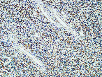
- Immunohistochemical analysis of paraffin-embedded Human Amygdala. 1, Antibody was diluted at 1:200(4° overnight). 2, High-pressure and temperature EDTA, pH8.0 was used for antigen retrieval. 3,Secondary antibody was diluted at 1:200(room temperature, 30min).
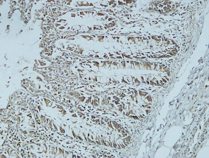
- Immunohistochemical analysis of paraffin-embedded Human colon. 1, Antibody was diluted at 1:400(4° overnight). 2, High-pressure and temperature EDTA, pH8.0 was used for antigen retrieval. 3,Secondary antibody was diluted at 1:200(room temperature, 30min).
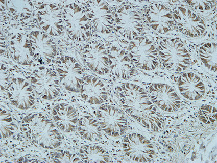
- Immunohistochemical analysis of paraffin-embedded Human colon. 1, Antibody was diluted at 1:400(4° overnight). 2, High-pressure and temperature EDTA, pH8.0 was used for antigen retrieval. 3,Secondary antibody was diluted at 1:200(room temperature, 30min).
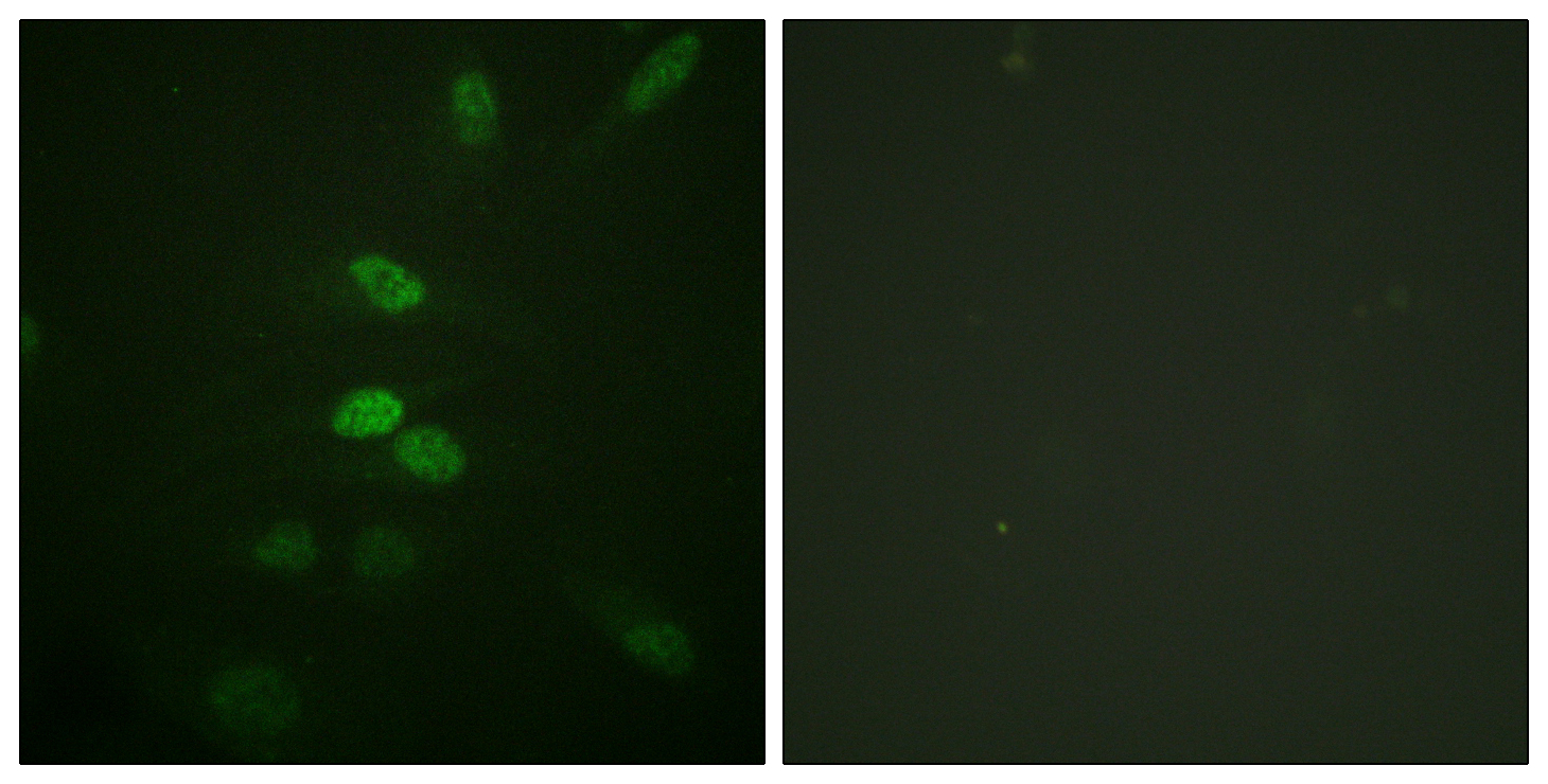
- Immunofluorescence analysis of HeLa cells, using CDC25B Antibody. The picture on the right is blocked with the synthesized peptide.
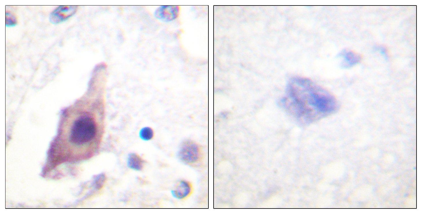
- Immunohistochemistry analysis of paraffin-embedded human brain tissue, using CDC25B Antibody. The picture on the right is blocked with the synthesized peptide.
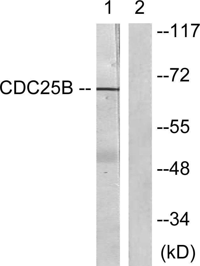
- Western blot analysis of lysates from Raw264.7 cells, treated with UV 15', using CDC25B Antibody. The lane on the right is blocked with the synthesized peptide.


