CD284 Polyclonal Antibody
- Catalog No.:YT0744
- Applications:IF;WB;IHC;ELISA
- Reactivity:Human;Mouse
- Target:
- CD284
- Fields:
- >>NF-kappa B signaling pathway;>>HIF-1 signaling pathway;>>Phagosome;>>PI3K-Akt signaling pathway;>>Necroptosis;>>Neutrophil extracellular trap formation;>>Toll-like receptor signaling pathway;>>NOD-like receptor signaling pathway;>>Alcoholic liver disease;>>Pathogenic Escherichia coli infection;>>Shigellosis;>>Salmonella infection;>>Pertussis;>>Legionellosis;>>Yersinia infection;>>Leishmaniasis;>>Chagas disease;>>Malaria;>>Toxoplasmosis;>>Amoebiasis;>>Tuberculosis;>>Hepatitis B;>>Measles;>>Influenza A;>>Human immunodeficiency virus 1 infection;>>Coronavirus disease - COVID-19;>>Proteoglycans in cancer;>>PD-L1 expression and PD-1 checkpoint pathway in cancer;>>Inflammatory bowel disease;>>Rheumatoid arthritis;>>Lipid and atherosclerosis
- Gene Name:
- TLR4
- Protein Name:
- Toll-like receptor 4
- Human Gene Id:
- 7099
- Human Swiss Prot No:
- O00206
- Mouse Gene Id:
- 21898
- Mouse Swiss Prot No:
- Q9QUK6
- Immunogen:
- The antiserum was produced against synthesized peptide derived from human CD284. AA range:392-441
- Specificity:
- CD284 Polyclonal Antibody detects endogenous levels of CD284 protein.
- Formulation:
- Liquid in PBS containing 50% glycerol, 0.5% BSA and 0.02% sodium azide.
- Source:
- Polyclonal, Rabbit,IgG
- Dilution:
- IF 1:50-200 WB 1:500 - 1:2000. IHC 1:100 - 1:300. ELISA: 1:40000. Not yet tested in other applications.
- Purification:
- The antibody was affinity-purified from rabbit antiserum by affinity-chromatography using epitope-specific immunogen.
- Concentration:
- 1 mg/ml
- Storage Stability:
- -15°C to -25°C/1 year(Do not lower than -25°C)
- Other Name:
- TLR4;Toll-like receptor 4;hToll;CD antigen CD284
- Observed Band(KD):
- 95kD
- Background:
- The protein encoded by this gene is a member of the Toll-like receptor (TLR) family which plays a fundamental role in pathogen recognition and activation of innate immunity. TLRs are highly conserved from Drosophila to humans and share structural and functional similarities. They recognize pathogen-associated molecular patterns that are expressed on infectious agents, and mediate the production of cytokines necessary for the development of effective immunity. The various TLRs exhibit different patterns of expression. This receptor has been implicated in signal transduction events induced by lipopolysaccharide (LPS) found in most gram-negative bacteria. Mutations in this gene have been associated with differences in LPS responsiveness. Multiple transcript variants encoding different isoforms have been found for this gene. [provided by RefSeq, Jan 2012],
- Function:
- disease:Genetic variation in TLR4 is associated with age-related macular degeneration type 10 (ARMD10) [MIM:611488]. ARMD is a multifactorial eye disease and the most common cause of irreversible vision loss in the developed world. In most patients, the disease is manifest as ophthalmoscopically visible yellowish accumulations of protein and lipid that lie beneath the retinal pigment epithelium and within an elastin-containing structure known as Bruch membrane.,domain:The TIR domain mediates interaction with NOX4.,function:Cooperates with LY96 and CD14 to mediate the innate immune response to bacterial lipopolysaccharide (LPS). Acts via MYD88, TIRAP and TRAF6, leading to NF-kappa-B activation, cytokine secretion and the inflammatory response.,polymorphism:Allele TLR4*B (Gly-299, Ile-399) is associated with a blunted response to inhaled LPS.,PTM:N-glycosylated. Glycosylation of Asn-526 an
- Subcellular Location:
- Cell membrane ; Single-pass type I membrane protein . Early endosome . Cell projection, ruffle . Upon complex formation with CD36 and TLR6, internalized through dynamin-dependent endocytosis (PubMed:20037584). Colocalizes with RFTN1 at cell membrane and then together with RFTN1 moves to endosomes, upon lipopolysaccharide stimulation. .
- Expression:
- Highly expressed in placenta, spleen and peripheral blood leukocytes (PubMed:9435236, PubMed:9237759). Detected in monocytes, macrophages, dendritic cells and several types of T-cells (PubMed:9237759, PubMed:27022195).
Ethyl pyruvate protects against Salmonella intestinal infection in mice through down-regulation of pro-inflammatory factors and inhibition of TLR4/MAPK pathway. INTERNATIONAL IMMUNOPHARMACOLOGY Int Immunopharmacol. 2019 Jun;71:155 WB Mouse jejunal
S100A8 facilitates the migration of colorectal cancer cells through regulating macrophages in the inflammatory microenvironment. ONCOLOGY REPORTS Oncol Rep. 2016 Jul;36(1):279-290 WB,IF Human 1:1000 THP-1 macrophages
Microparticulated Polygonatum sibiricum polysaccharide shows potent vaccine adjuvant effect INTERNATIONAL JOURNAL OF PHARMACEUTICS Kai Shen WB Mouse 1:250 bone marrow-derived dendritic cell (BMDC)
Galectin from Trichinella spiralis alleviates DSS-induced colitis in mice by regulating the intestinal microbiota VETERINARY RESEARCH Li Jianqing WB Mouse 1:1000 colon tissue
- June 19-2018
- WESTERN IMMUNOBLOTTING PROTOCOL
- June 19-2018
- IMMUNOHISTOCHEMISTRY-PARAFFIN PROTOCOL
- June 19-2018
- IMMUNOFLUORESCENCE PROTOCOL
- September 08-2020
- FLOW-CYTOMEYRT-PROTOCOL
- May 20-2022
- Cell-Based ELISA│解您多样本WB检测之困扰
- July 13-2018
- CELL-BASED-ELISA-PROTOCOL-FOR-ACETYL-PROTEIN
- July 13-2018
- CELL-BASED-ELISA-PROTOCOL-FOR-PHOSPHO-PROTEIN
- July 13-2018
- Antibody-FAQs
- Products Images
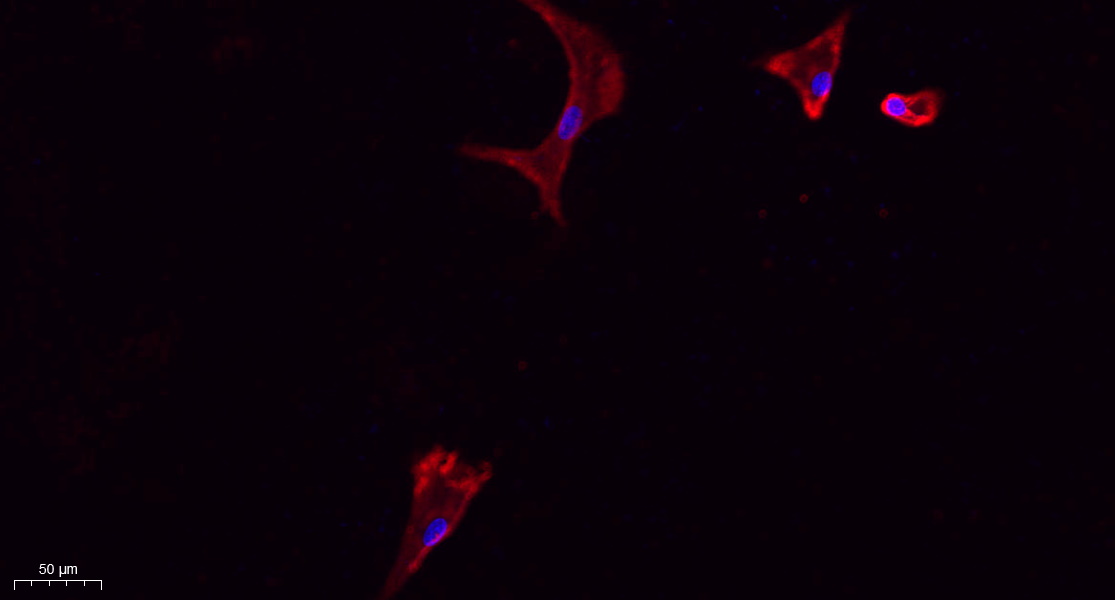
- Immunofluorescence analysis of A549. 1,primary Antibody(red) was diluted at 1:200(4°C overnight). 2, Goat Anti Rabbit IgG (H&L) - Alexa Fluor 594 Secondary antibody was diluted at 1:1000(room temperature, 50min).3, Picture B: DAPI(blue) 10min.
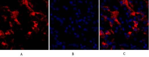
- Immunofluorescence analysis of human-stomach tissue. 1,CD284 Polyclonal Antibody(red) was diluted at 1:200(4°C,overnight). 2, Cy3 labled Secondary antibody was diluted at 1:300(room temperature, 50min).3, Picture B: DAPI(blue) 10min. Picture A:Target. Picture B: DAPI. Picture C: merge of A+B
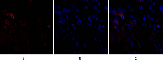
- Immunofluorescence analysis of mouse-liver tissue. 1,CD284 Polyclonal Antibody(red) was diluted at 1:200(4°C,overnight). 2, Cy3 labled Secondary antibody was diluted at 1:300(room temperature, 50min).3, Picture B: DAPI(blue) 10min. Picture A:Target. Picture B: DAPI. Picture C: merge of A+B
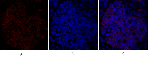
- Immunofluorescence analysis of mouse-spleen tissue. 1,CD284 Polyclonal Antibody(red) was diluted at 1:200(4°C,overnight). 2, Cy3 labled Secondary antibody was diluted at 1:300(room temperature, 50min).3, Picture B: DAPI(blue) 10min. Picture A:Target. Picture B: DAPI. Picture C: merge of A+B
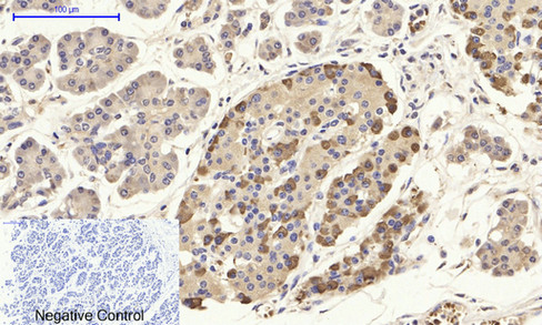
- Immunohistochemical analysis of paraffin-embedded Human-stomach-cancer tissue. 1,CD284 Polyclonal Antibody was diluted at 1:200(4°C,overnight). 2, Sodium citrate pH 6.0 was used for antibody retrieval(>98°C,20min). 3,Secondary antibody was diluted at 1:200(room tempeRature, 30min). Negative control was used by secondary antibody only.
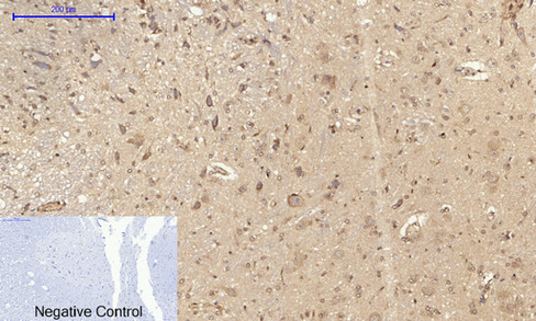
- Immunohistochemical analysis of paraffin-embedded Rat-spinal-cord tissue. 1,CD284 Polyclonal Antibody was diluted at 1:200(4°C,overnight). 2, Sodium citrate pH 6.0 was used for antibody retrieval(>98°C,20min). 3,Secondary antibody was diluted at 1:200(room tempeRature, 30min). Negative control was used by secondary antibody only.
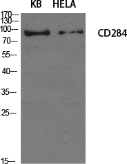
- Western Blot analysis of various cells using CD284 Polyclonal Antibody diluted at 1:1000
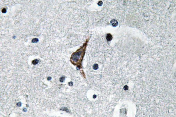
- Immunohistochemistry analysis of TLR4 antibody in paraffin-embedded human brain tissue.

- Western blot analysis of lysate from HeLa cells, using TLR4 antibody.


