Caveolin-1 Polyclonal Antibody
- Catalog No.:YT0686
- Applications:WB;IHC;IF;ELISA
- Reactivity:Human;Mouse;Rat
- Target:
- Caveolin-1
- Fields:
- >>Endocytosis;>>Focal adhesion;>>Prion disease;>>Bacterial invasion of epithelial cells;>>Proteoglycans in cancer;>>Viral myocarditis;>>Fluid shear stress and atherosclerosis
- Gene Name:
- CAV1
- Protein Name:
- Caveolin-1
- Human Gene Id:
- 857
- Human Swiss Prot No:
- Q03135
- Mouse Gene Id:
- 12389
- Mouse Swiss Prot No:
- P49817
- Rat Swiss Prot No:
- P41350
- Immunogen:
- The antiserum was produced against synthesized peptide derived from human Caveolin-1. AA range:129-178
- Specificity:
- Caveolin-1 Polyclonal Antibody detects endogenous levels of Caveolin-1 protein.
- Formulation:
- Liquid in PBS containing 50% glycerol, 0.5% BSA and 0.02% sodium azide.
- Source:
- Polyclonal, Rabbit,IgG
- Dilution:
- WB 1:500-2000, IF 1:50-300, IHC 1:50-300
- Purification:
- The antibody was affinity-purified from rabbit antiserum by affinity-chromatography using epitope-specific immunogen.
- Concentration:
- 1 mg/ml
- Storage Stability:
- -15°C to -25°C/1 year(Do not lower than -25°C)
- Other Name:
- CAV1;CAV;Caveolin-1
- Observed Band(KD):
- 25kD
- Background:
- The scaffolding protein encoded by this gene is the main component of the caveolae plasma membranes found in most cell types. The protein links integrin subunits to the tyrosine kinase FYN, an initiating step in coupling integrins to the Ras-ERK pathway and promoting cell cycle progression. The gene is a tumor suppressor gene candidate and a negative regulator of the Ras-p42/44 mitogen-activated kinase cascade. Caveolin 1 and caveolin 2 are located next to each other on chromosome 7 and express colocalizing proteins that form a stable hetero-oligomeric complex. Mutations in this gene have been associated with Berardinelli-Seip congenital lipodystrophy. Alternatively spliced transcripts encode alpha and beta isoforms of caveolin 1.[provided by RefSeq, Mar 2010],
- Function:
- disease:Defects in CAV1 are the cause of congenital generalized lipodystrophy type 3 (CGL3) [MIM:612526]; also called Berardinelli-Seip congenital lipodystrophy type 3 (BSCL3). Congenital generalized lipodystrophies are autosomal recessive disorders characterized by a near absence of adipose tissue, extreme insulin resistance, hypertriglyceridemia, hepatic steatosis and early onset of diabetes.,function:May act as a scaffolding protein within caveolar membranes. Interacts directly with G-protein alpha subunits and can functionally regulate their activity.,online information:Caveolin entry,PTM:The initiator methionine for isoform Beta is removed during or just after translation. The new N-terminal amino acid is then N-acetylated.,similarity:Belongs to the caveolin family.,subcellular location:Potential hairpin-like structure in the membrane. Membrane protein of caveolae.,subunit:Homooligo
- Subcellular Location:
- Golgi apparatus membrane; Peripheral membrane protein. Cell membrane; Peripheral membrane protein. Membrane, caveola ; Peripheral membrane protein. Membrane raft . Golgi apparatus, trans-Golgi network . Colocalized with DPP4 in membrane rafts. Potential hairpin-like structure in the membrane. Membrane protein of caveolae.
- Expression:
- Skeletal muscle, liver, stomach, lung, kidney and heart (at protein level). Expressed in the brain.
Graphene and graphene oxide as a solid matrix for extraction of membrane and membrane-associated proteins. MICROCHIMICA ACTA 2018 Jan 19 WB Human 95D cell,BEAS-2B cell
Novel peptide GX1 inhibits angiogenesis by specifically binding to transglutaminase-2 in the tumorous endothelial cells of gastric cancer. Cell Death & Disease Cell Death Dis. 2018 May;9(6):1-16 WB Human 1:500 HUVECs,SGC7901 cell,GES cell
Caveolin-1 affects tumor drug resistance in esophageal squamous cell carcinoma by regulating expressions of P-gp and MRP1. TUMOR BIOLOGY Tumor Biol. 2016 Jul;37(7):9189-9196 WB,IHC Human 1:100 esophageal squamous cell carcinoma tissues Ec9706 cell
Uzzaman, Asad, et al. "Graphene and graphene oxide as a solid matrix for extraction of membrane and membrane-associated proteins." Microchimica Acta 185.2 (2018): 123.
- June 19-2018
- WESTERN IMMUNOBLOTTING PROTOCOL
- June 19-2018
- IMMUNOHISTOCHEMISTRY-PARAFFIN PROTOCOL
- June 19-2018
- IMMUNOFLUORESCENCE PROTOCOL
- September 08-2020
- FLOW-CYTOMEYRT-PROTOCOL
- May 20-2022
- Cell-Based ELISA│解您多样本WB检测之困扰
- July 13-2018
- CELL-BASED-ELISA-PROTOCOL-FOR-ACETYL-PROTEIN
- July 13-2018
- CELL-BASED-ELISA-PROTOCOL-FOR-PHOSPHO-PROTEIN
- July 13-2018
- Antibody-FAQs
- Products Images
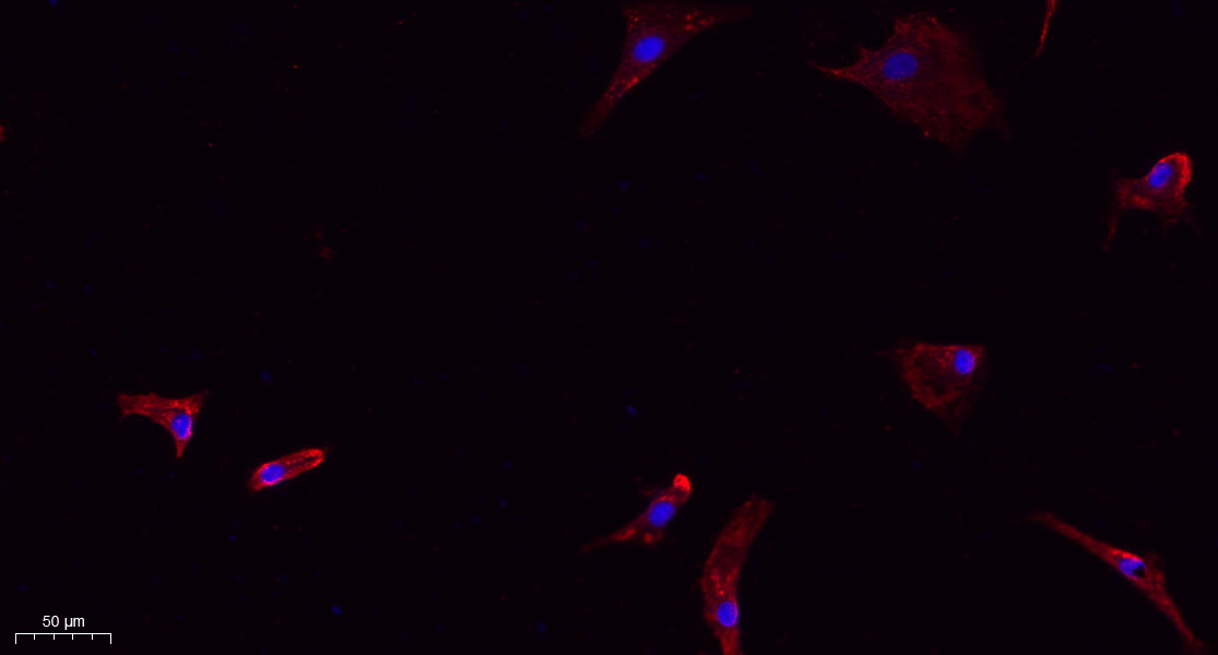
- Immunofluorescence analysis of A549. 1,primary Antibody(red) was diluted at 1:200(4°C overnight). 2, Goat Anti Rabbit IgG (H&L) - Alexa Fluor 594 Secondary antibody was diluted at 1:1000(room temperature, 50min).3, Picture B: DAPI(blue) 10min.
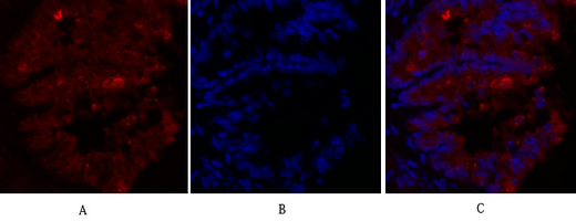
- Immunofluorescence analysis of rat-lung tissue. 1,Caveolin-1 Polyclonal Antibody(red) was diluted at 1:200(4°C,overnight). 2, Cy3 labled Secondary antibody was diluted at 1:300(room temperature, 50min).3, Picture B: DAPI(blue) 10min. Picture A:Target. Picture B: DAPI. Picture C: merge of A+B
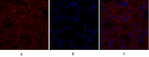
- Immunofluorescence analysis of rat-kidney tissue. 1,Caveolin-1 Polyclonal Antibody(red) was diluted at 1:200(4°C,overnight). 2, Cy3 labled Secondary antibody was diluted at 1:300(room temperature, 50min).3, Picture B: DAPI(blue) 10min. Picture A:Target. Picture B: DAPI. Picture C: merge of A+B
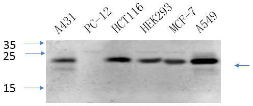
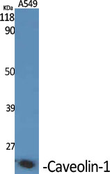
- Western Blot analysis of various cells using primary antibody diluted at 1:1000(4°C overnight). Secondary antibody:Goat Anti-rabbit IgG IRDye 800( diluted at 1:5000, 25°C, 1 hour). Cell lysate was extracted by Minute™ Plasma Membrane Protein Isolation and Cell Fractionation Kit(SM-005, Inventbiotech,MN,USA).
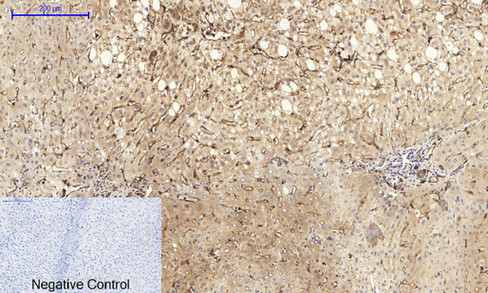
- Immunohistochemical analysis of paraffin-embedded Human-liver tissue. 1,Caveolin-1 Polyclonal Antibody was diluted at 1:200(4°C,overnight). 2, Sodium citrate pH 6.0 was used for antibody retrieval(>98°C,20min). 3,Secondary antibody was diluted at 1:200(room tempeRature, 30min). Negative control was used by secondary antibody only.
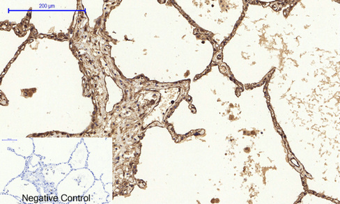
- Immunohistochemical analysis of paraffin-embedded Human-lung tissue. 1,Caveolin-1 Polyclonal Antibody was diluted at 1:200(4°C,overnight). 2, Sodium citrate pH 6.0 was used for antibody retrieval(>98°C,20min). 3,Secondary antibody was diluted at 1:200(room tempeRature, 30min). Negative control was used by secondary antibody only.
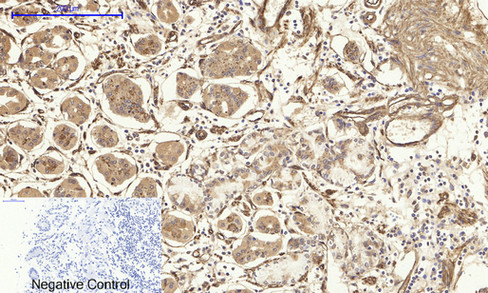
- Immunohistochemical analysis of paraffin-embedded Human-stomach tissue. 1,Caveolin-1 Polyclonal Antibody was diluted at 1:200(4°C,overnight). 2, Sodium citrate pH 6.0 was used for antibody retrieval(>98°C,20min). 3,Secondary antibody was diluted at 1:200(room tempeRature, 30min). Negative control was used by secondary antibody only.
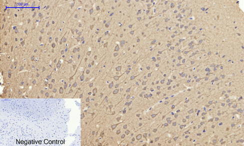
- Immunohistochemical analysis of paraffin-embedded Rat-brain tissue. 1,Caveolin-1 Polyclonal Antibody was diluted at 1:200(4°C,overnight). 2, Sodium citrate pH 6.0 was used for antibody retrieval(>98°C,20min). 3,Secondary antibody was diluted at 1:200(room tempeRature, 30min). Negative control was used by secondary antibody only.
- Western Blot analysis of various cells using Caveolin-1 Polyclonal Antibody diluted at 1:1000
.jpg)
- Western Blot analysis of HuvEc cells using Caveolin-1 Polyclonal Antibody diluted at 1:1000
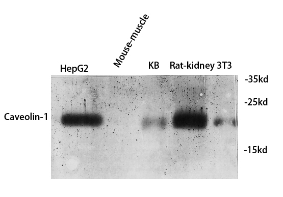
- Western blot analysis of various cell Lysate, antibody was diluted at 1:1000. Secondary antibody(catalog#:RS0002) was diluted at 1:20000
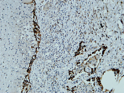
- Immunohistochemical analysis of paraffin-embedded Human lung. 1, Antibody was diluted at 1:200(4° overnight). 2, High-pressure and temperature EDTA, pH8.0 was used for antigen retrieval. 3,Secondary antibody was diluted at 1:200(room temperature, 30min).
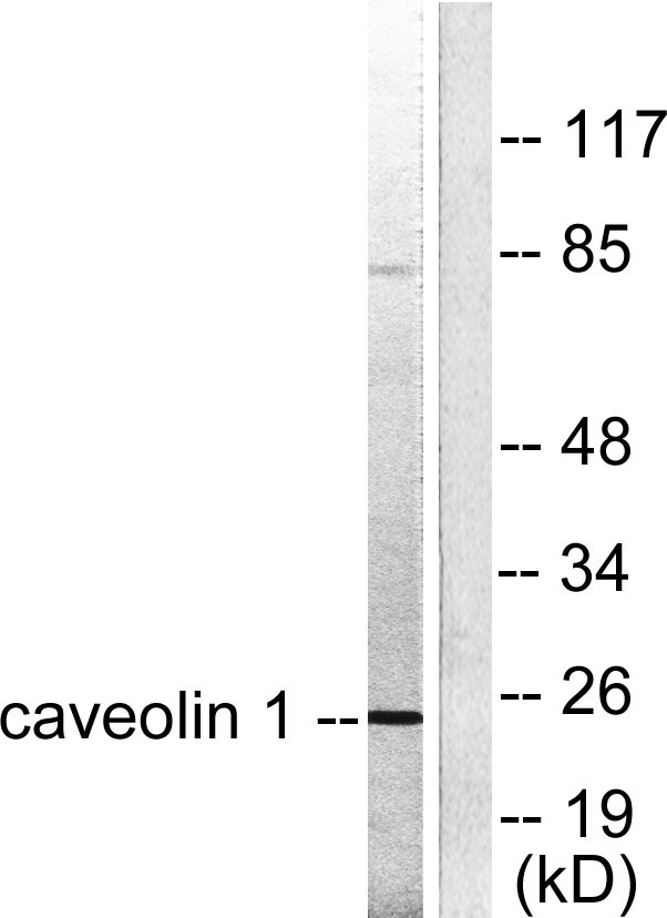
- Western blot analysis of lysates from HUVEC cells, using Caveolin-1 Antibody. The lane on the right is blocked with the synthesized peptide.


