Cadherin-16 Polyclonal Antibody
- Catalog No.:YT0594
- Applications:WB;IHC;IF;ELISA
- Reactivity:Human;Mouse
- Target:
- Cadherin-16
- Gene Name:
- CDH16
- Protein Name:
- Cadherin-16
- Human Gene Id:
- 1014
- Human Swiss Prot No:
- O75309
- Mouse Swiss Prot No:
- O88338
- Immunogen:
- The antiserum was produced against synthesized peptide derived from human CDH16. AA range:780-829
- Specificity:
- Cadherin-16 Polyclonal Antibody detects endogenous levels of Cadherin-16 protein.
- Formulation:
- Liquid in PBS containing 50% glycerol, 0.5% BSA and 0.02% sodium azide.
- Source:
- Polyclonal, Rabbit,IgG
- Dilution:
- WB 1:500 - 1:2000. IHC 1:100 - 1:300. IF 1:200 - 1:1000. ELISA: 1:10000. Not yet tested in other applications.
- Purification:
- The antibody was affinity-purified from rabbit antiserum by affinity-chromatography using epitope-specific immunogen.
- Concentration:
- 1 mg/ml
- Storage Stability:
- -15°C to -25°C/1 year(Do not lower than -25°C)
- Other Name:
- CDH16;Cadherin-16;Kidney-specific cadherin;Ksp-cadherin
- Observed Band(KD):
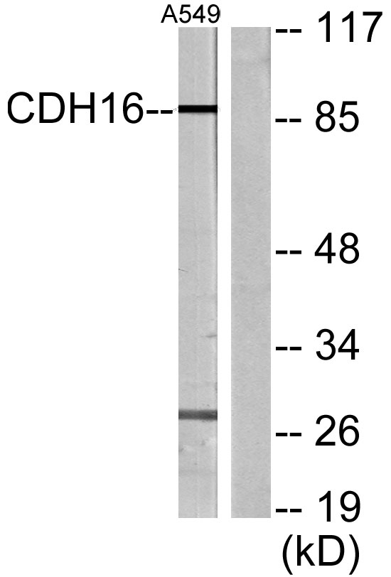
- 90kD
- Background:
- This gene is a member of the cadherin superfamily, genes encoding calcium-dependent, membrane-associated glycoproteins. Mapped to a previously identified cluster of cadherin genes on chromosome 16q22.1, the gene localizes with superfamily members CDH1, CDH3, CDH5, CDH8 and CDH11. The protein consists of an extracellular domain containing 6 cadherin domains, a transmembrane region and a truncated cytoplasmic domain but lacks the prosequence and tripeptide HAV adhesion recognition sequence typical of most classical cadherins. Expression is exclusively in kidney, where the protein functions as the principal mediator of homotypic cellular recognition, playing a role in the morphogenic direction of tissue development. Alternatively spliced transcript variants encoding distinct isoforms have been identified. [provided by RefSeq, Mar 2011],
- Function:
- function:Cadherins are calcium dependent cell adhesion proteins. They preferentially interact with themselves in a homophilic manner in connecting cells; cadherins may thus contribute to the sorting of heterogeneous cell types.,similarity:Contains 6 cadherin domains.,tissue specificity:Kidney specific.,
- Subcellular Location:
- Cell membrane ; Single-pass type I membrane protein .
- Expression:
- Kidney specific.
- June 19-2018
- WESTERN IMMUNOBLOTTING PROTOCOL
- June 19-2018
- IMMUNOHISTOCHEMISTRY-PARAFFIN PROTOCOL
- June 19-2018
- IMMUNOFLUORESCENCE PROTOCOL
- September 08-2020
- FLOW-CYTOMEYRT-PROTOCOL
- May 20-2022
- Cell-Based ELISA│解您多样本WB检测之困扰
- July 13-2018
- CELL-BASED-ELISA-PROTOCOL-FOR-ACETYL-PROTEIN
- July 13-2018
- CELL-BASED-ELISA-PROTOCOL-FOR-PHOSPHO-PROTEIN
- July 13-2018
- Antibody-FAQs
- Products Images

- Western Blot analysis of various cells using Cadherin-16 Polyclonal Antibody diluted at 1:1000
.jpg)
- Western Blot analysis of A549 cells using Cadherin-16 Polyclonal Antibody diluted at 1:1000

- Western blot analysis of lysates from A549 cells, using CDH16 Antibody. The lane on the right is blocked with the synthesized peptide.

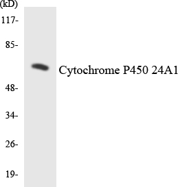
- Western blot analysis of the lysates from HepG2 cells using Cytochrome P450 24A1 antibody.

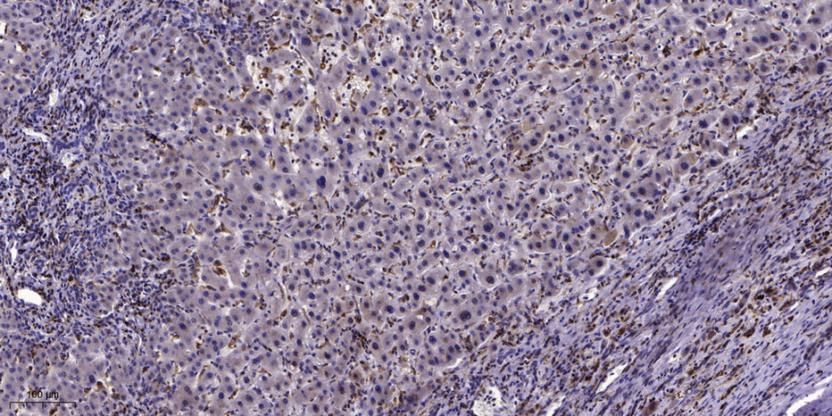
- Immunohistochemical analysis of paraffin-embedded human liver cancer. 1, Antibody was diluted at 1:200(4° overnight). 2, Tris-EDTA,pH9.0 was used for antigen retrieval. 3,Secondary antibody was diluted at 1:200(room temperature, 45min).


